

Corneal pachymetry
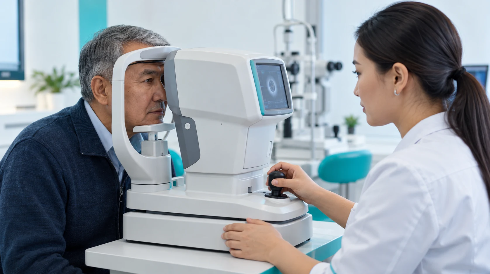
Pachymetry measures corneal thickness.
Learn moreSoft contact lenses can temporarily alter the shape of the cornea and distort measurement results. Therefore, they should be removed 2-3 days prior to the examination.
If pharmacological pupil dilation is planned, you may experience light sensitivity and blurred near vision for 3 to 4 hours afterward.
Pachymetry measures corneal thickness.
Learn moreTonometry measures intraocular pressure using a contact or non-contact method.
Learn moreOCT produces cross-sectional images of eye structures without surgery.
Learn morePerimetry tests the visual field and helps identify areas of reduced sensitivity.
Learn moreA preoperative examination assesses the safety of laser vision correction and helps select a suitable method. It evaluates refraction, corneal shape and thickness, the ocular surface and other eye structures.
Who is this assessment for? For patients with myopia, hyperopia or astigmatism who want to understand whether laser vision correction can safely be considered and which approach should be discussed with a refractive surgeon.
What symptoms or goals commonly lead to assessment? blurred distance vision; blurred near vision; myopia; hyperopia; astigmatism; dependence on glasses; dependence on contact lenses; poor contact-lens tolerance; glare or halos in dim light; a desire to reduce dependence on optical correction.
Age and presbyopia After about age 40, near vision and the possible future need for reading correction should be discussed separately. Laser correction of refractive error does not stop the natural age-related changes in the eye's ability to focus at near. A good distance-vision result therefore does not mean that the patient will never need reading glasses.
The doctor may defer a final decision for unstable refraction, active inflammation, marked dry eye, an unstable ocular surface, suspected keratoconus or ectasia, an uncertain corneal map, retinal changes or another eye condition requiring treatment or further diagnostics.
Treatment, observation or repeat measurements come first.
Diagnostics do not guarantee a specific postoperative acuity.
An unstable ocular surface can reduce corneal measurement reliability.
Suspected keratoconus or ectasia needs separate assessment.
A thin cornea does not automatically exclude correction, but requires comprehensive assessment; nor does it automatically establish suitability for Femto LASIK, PRK or another method.
Peripheral retinal changes do not automatically require prophylactic laser. A specialist decides on specific indications.
After drug-induced pupil dilation, light sensitivity and blurred near vision may last 3–4 hours.
An inadequate lens break may distort topography or tomography. Soft contact lenses may temporarily alter corneal shape and distort measurements. They should therefore be removed 2–3 days before diagnostics.
After dilation, do not drive until vision and light sensitivity return to a comfortable level.
What is particularly important to tell the doctor? How your glasses or contact lens prescription has changed in recent years; which contact lenses you use; whether you have worn orthokeratology lenses; any dryness, burning or gritty sensation; previous eye injuries or operations; corneal disease; retinal disease; glaucoma; chronic and systemic conditions; and all regularly used medicines.
Urgent ophthalmic assessment is more appropriate than routine refractive-surgery screening if there is: sudden significant vision loss; severe eye pain; marked redness; a curtain-like shadow; a sudden increase in floaters; new flashes of light; eye trauma.
Diagnostics do not guarantee surgical eligibility.
Book an eye examination
The doctor clarifies symptoms, the history of refractive changes, contact lens type and wearing schedule, previous diseases and operations, medications, occupation, sports and expectations of laser vision correction.
Takes: 15 min

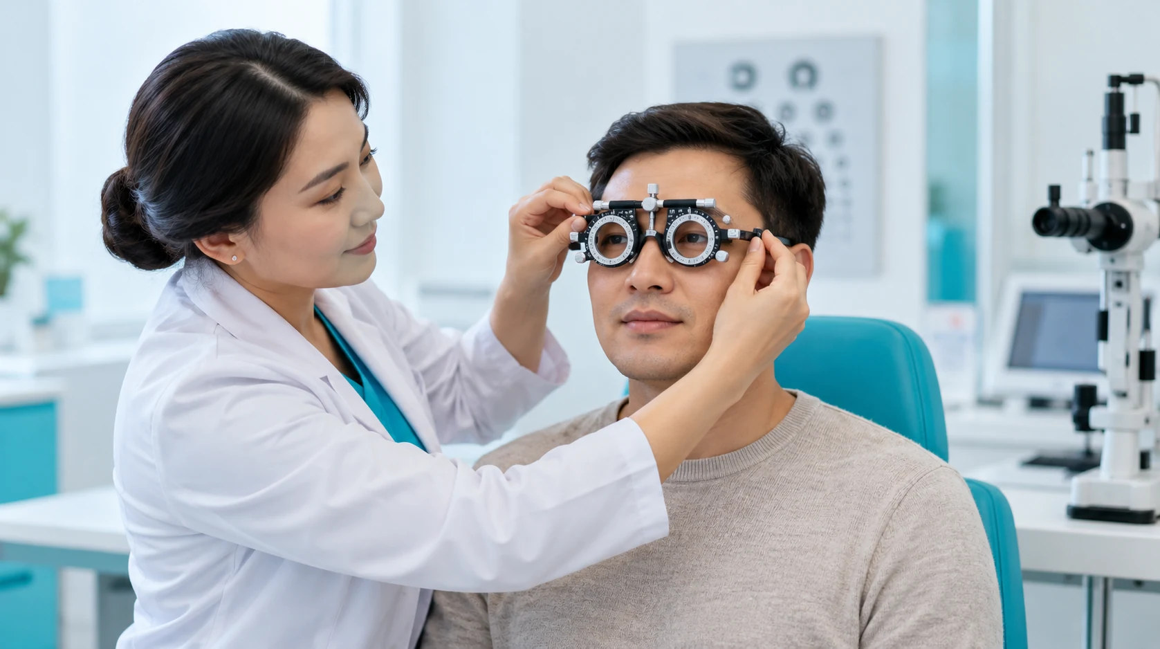
Vision without and with optimal correction, and objective and subjective refraction, are assessed.
Takes: 10 min
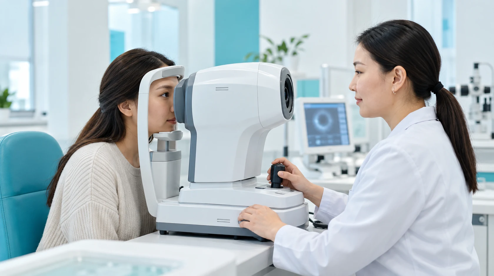
Maps of the anterior and posterior corneal surfaces and thickness distribution are obtained.
Takes: 5 min
Pachymetry and additional measurements assess corneal thickness and anterior-segment anatomy in relation to the proposed correction method.
Takes: 5 min
Biomicroscopy and tear-film assessment help detect dryness, inflammation, eyelid and corneal disease and other conditions affecting measurement accuracy and postoperative comfort.
Takes: 5 min
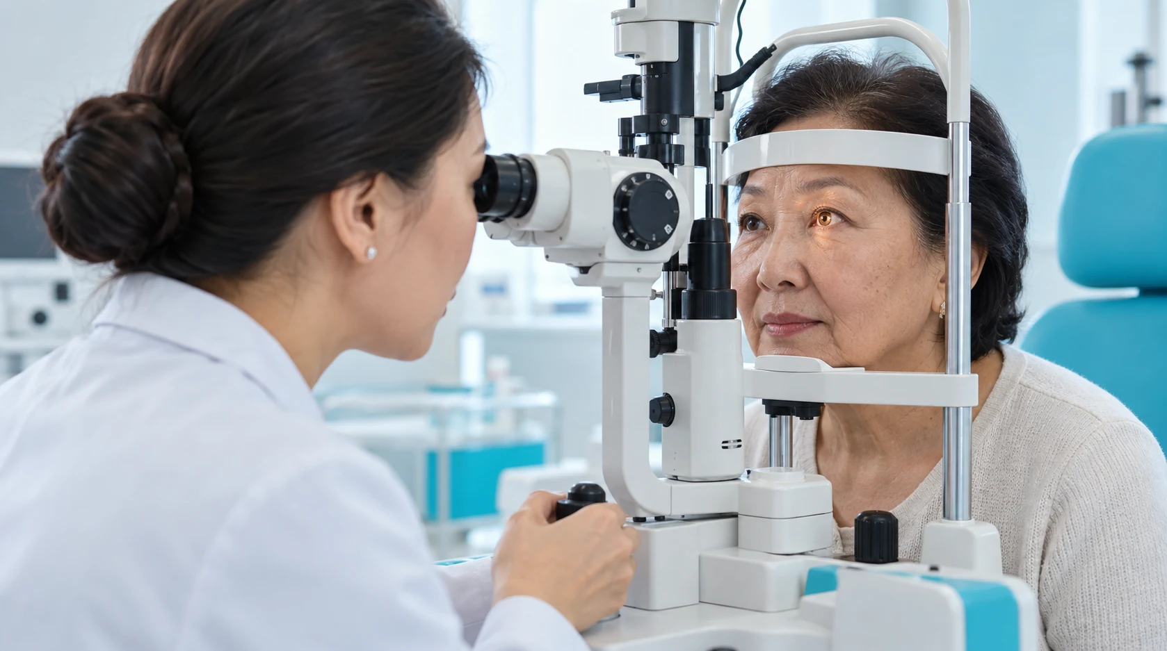
Tonometry assesses intraocular pressure.
Takes: 5 min
The pupil may be dilated to assess the retina, macula and optic disc according to protocol and indications.
Takes: 30 - 40 min
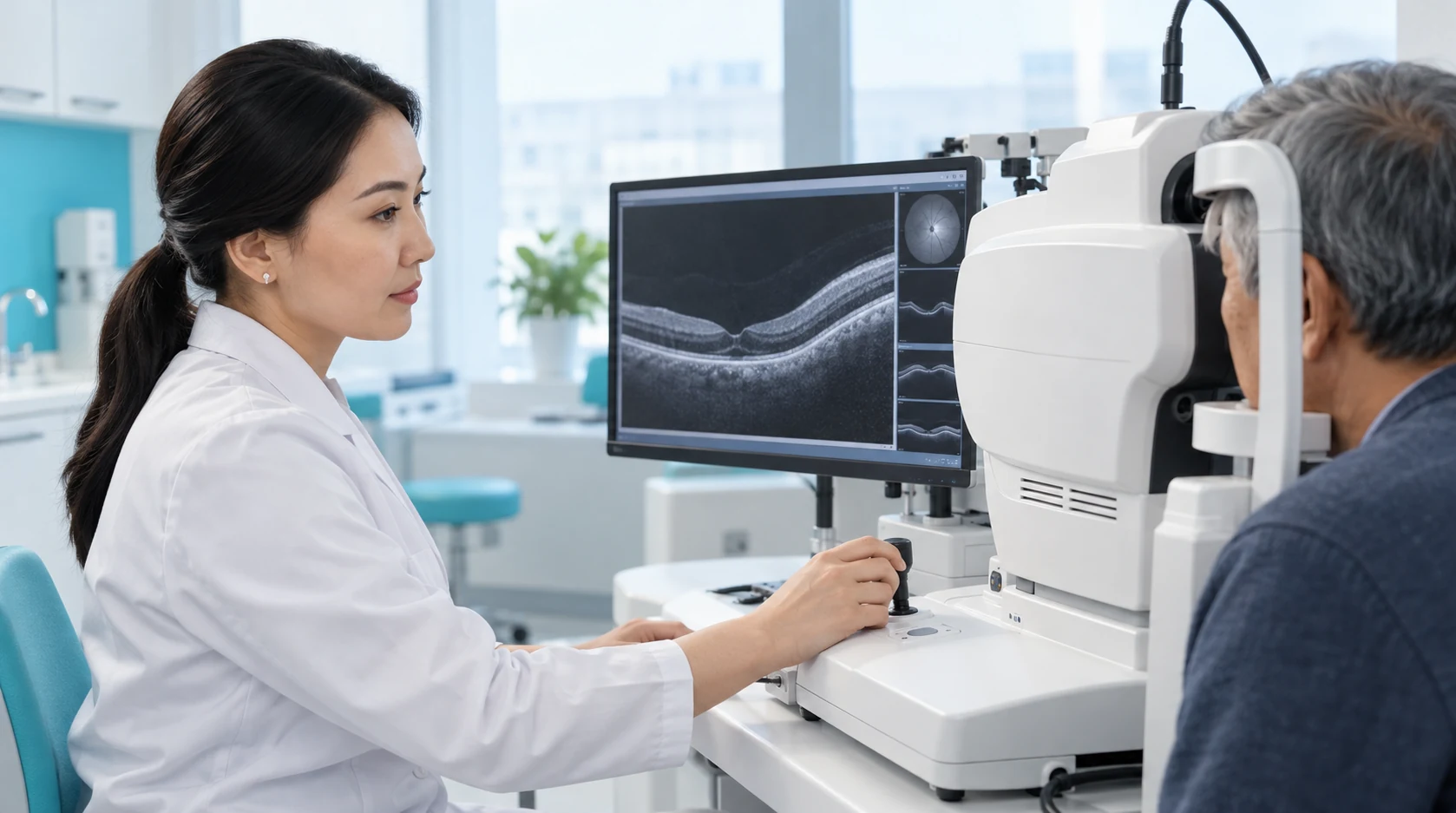
The doctor integrates all results, discusses candidacy, limitations and alternatives, explains realistic expectations and provides a next-step plan.
Takes: 20 min
Diagnostics help detect ectasia signs and other findings that may influence the choice of procedure.
Corneal and refractive measurements help the surgeon assess suitability and plan the selected laser technique.
Ocular surface assessment helps identify dryness and address it before final calculations when needed.
Before intervention, the doctor discusses visual goals, limitations and possible need for glasses afterwards.
Book an examination so the doctor can assess vision and corneal measurements, exclude significant limitations and explain correction methods or alternatives. Book an assessment
A team of experienced doctors across specialties.

Candidate of Medical Sciences; Highest-category ophthalmologist

Highest-category ophthalmologist

Second-category ophthalmologist

Second-category ophthalmologist


A routine test may establish how well a person sees and which optical correction is needed.
Preoperative refractive assessment asks more complex questions: is refraction stable, is the cornea suitable for the procedure, are there signs of ectasia or ocular-surface disease, what is best-corrected acuity, and are there associated findings that alter treatment?
The assessment should explain not only eligibility but why, which methods can be discussed, which restrictions remain and whether ocular-surface, retinal or other disease needs treatment first.
Diagnostic equipment supplies measurements but does not automatically select surgery. The decision follows comparison of all findings, the capabilities of the particular technology and the patient's visual needs.
The ophthalmologist correlates refraction and visual acuity with corneal topography or tomography and thickness, ocular surface status, intraocular pressure and retinal findings.
The outcome is not a promise of surgery, but a medical conclusion: whether laser vision correction can be considered, which methods should be discussed, which limitations must be taken into account, and whether additional steps are needed before treatment.
Vision without and with optimal correction, and objective and subjective refraction, are assessed.
The doctor assesses refraction after cycloplegia when needed to reduce the effect of accommodation.
Maps of the anterior and posterior corneal surfaces and thickness distribution are obtained.
This is a key screening stage for keratoconus, other ectasias, irregular astigmatism and lens-related corneal shape changes.
Tonometry assesses intraocular pressure.
Abnormal findings may require further diagnostics to exclude or assess glaucoma.
The pupil may be dilated to assess the retina, macula and optic disc according to protocol and indications.
A specialist determines further management if suspicious changes are found.
Discontinue contact lenses for the interval set by KazRIED.
Bring:
Dryness, blepharitis or inflammation is treated first.
Key corneal measurements may be repeated after surface stabilisation.
The doctor may recommend:
The patient may be referred to a retinal specialist or laser surgeon.
Additional treatment is given only for medical indications.
The doctor explains why and, when indicated, may discuss:
After clearance, the patient receives separate KazRIED instructions covering:
The diagnostic package includes Pentacam, Corvis ST, Schwind Sirius, cycloscopy and a doctor's consultation. Additional tests indicated clinically are charged separately.
The value lies in the doctor's interpretation of all findings, not the number of devices or measurements.
A good report answers specific questions:
Related information: Contact Lenses for Children.
Kazakhstan’s first specialized ophthalmology institute, operating since 1933.
Modern diagnostics and surgery using world-class equipment.
Leading ophthalmologists in Kazakhstan, scientists and experienced surgeons.
More than 30,000 operations and around 270,000 consultations annually.


Kazakhstan’s first specialized ophthalmology institute, operating since 1933.
Modern diagnostics and surgery using world-class equipment.
Leading ophthalmologists in Kazakhstan, scientists and experienced surgeons.
More than 30,000 operations and around 270,000 consultations annually.
Kazakhstan’s first specialized ophthalmology institute, operating since 1933.
Modern diagnostics and surgery using world-class equipment.
Leading ophthalmologists in Kazakhstan, scientists and experienced surgeons.
More than 30,000 operations and around 270,000 consultations annually.


