
Laser glaucoma treatment
Laser treatment lowers eye pressure through different mechanisms.
Learn moreLaser treatment lowers eye pressure through different mechanisms.
Learn moreTrabeculectomy creates an additional route for aqueous outflow to lower eye pressure.
Learn moreGlaucoma drainage procedures improve aqueous outflow and lower eye pressure.
Learn moreGlaucoma treatment aims to lower intraocular pressure and preserve remaining visual function. Depending on disease stage and progression risk, care may involve drops, laser treatment or surgery.
If glaucoma has already been diagnosed, treatment needs to be selected or reviewed and intraocular pressure, visual field and optic nerve status monitored regularly.
Raised intraocular pressure (IOP) alone does not always mean glaucoma is present. However, it requires a comprehensive assessment of the optic nerve and other risk factors to determine whether monitoring or treatment is needed.
Progressive changes in the visual field, optic disc or nerve fibre layer may require more intensive treatment even without subjective symptoms.
If target IOP is not reached, the disease continues to progress, medicines are poorly tolerated or the drop regimen is difficult to follow, the doctor may consider laser or surgical treatment.
In open-angle glaucoma, the anterior chamber angle remains open, but fluid drainage through the eye’s drainage system is impaired or insufficient. The disease often develops gradually and can remain without obvious symptoms for a long time.
In angle-closure glaucoma, the iris partly or completely blocks the anterior chamber angle, preventing normal drainage of fluid. The disease may develop gradually, but an acute attack causes a sharp rise in IOP and requires emergency medical attention.
Raised IOP and optic nerve damage develop as a result of another eye disease, trauma, inflammation, vascular changes or other causes.
Rare forms requiring a separate assessment and treatment pathway with a paediatric ophthalmologist and ophthalmic surgeon.
Children with suspected congenital or juvenile glaucoma undergo specialist assessment by a paediatric ophthalmologist.
When the diagnosis is confirmed, necessary surgical treatment for children is provided free of charge under compulsory social health insurance, in accordance with applicable rules and treatment indications.
Treatment is planned individually after a comprehensive ophthalmic examination.
Glaucoma requires long-term, regular follow-up. Lowering intraocular pressure does not mean that the disease is cured: pressure, visual fields and the optic nerve still need monitoring after successful laser or surgical treatment. Existing glaucomatous optic nerve damage and the associated visual loss are generally irreversible.
The doctor measures pressure, assesses the optic nerve, field and anterior-chamber angle and requests further tests if needed, to confirm diagnosis, type and stage and establish baseline measurements.
Disease stage, current pressure, progression rate, age and risk factors determine the target pressure and treatment strategy.
Severe eye pain, sudden blurred vision, a red eye, coloured halos around lights, marked headache, nausea or vomiting may indicate an acute attack of angle-closure glaucoma. This requires urgent ophthalmic care and must not wait for a routine appointment.
KazRIED is not an emergency medical service. If an acute attack of angle-closure glaucoma is suspected, the patient must immediately contact emergency medical services or the nearest city hospital providing emergency ophthalmic care.
Once the condition is stabilised and the acute attack is controlled, the patient may be referred to KazRIED for specialist assessment and further treatment planning.
Glaucoma requires long-term regular follow-up. Lower pressure does not mean cure: pressure, visual fields and the optic nerve need monitoring after successful laser or surgery. Existing glaucomatous damage and visual loss are generally irreversible. Treatment therefore aims to prevent or slow further progression and preserve remaining vision.
Book a consultation
The doctor measures pressure, assesses the optic nerve, visual field and anterior-chamber angle, and orders further tests when needed.
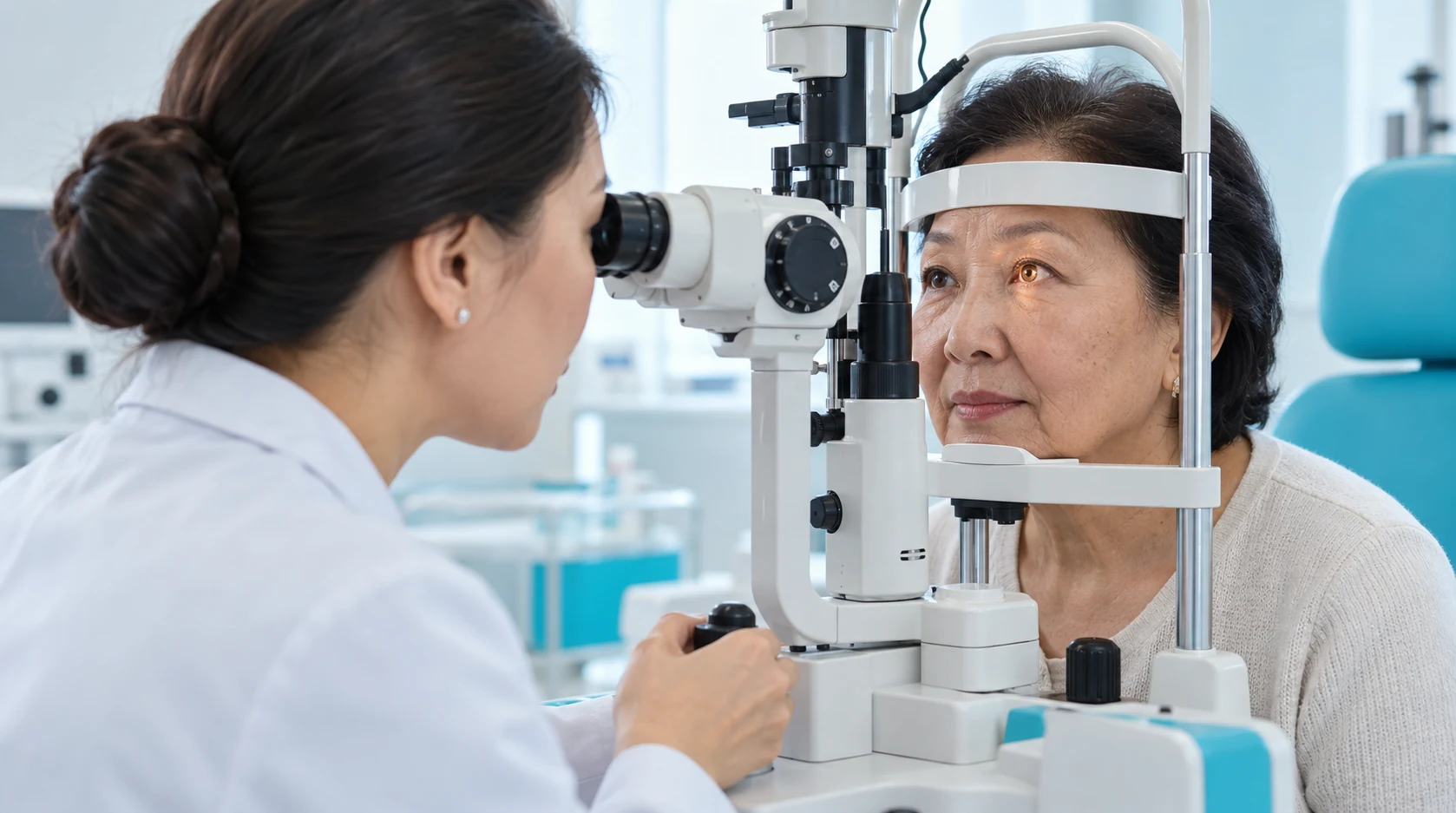
Disease stage, current pressure, progression rate, age and risk factors determine the target pressure and treatment strategy.

Duration depends on the method.
After starting or changing treatment, the doctor assesses pressure, tolerability and eye health.

Follow-up is ongoing.
After glaucoma laser treatment, review is at one week, then according to indications and the individual follow-up plan.
Reaching the target pressure helps reduce the risk of further optic nerve damage.
Treatment aims to protect remaining visual function; function already lost to glaucoma is usually not restored.
Drops, laser and surgery are selected according to glaucoma type, pressure and optic nerve health.
Repeat pressure measurements, visual field testing and optic nerve assessment help evaluate treatment effectiveness.
Start with a glaucoma assessment The doctor will assess intraocular pressure, the optic nerve and visual field, determine the form and stage of the disease and suggest a treatment or monitoring plan.
A team of experienced doctors across specialties.

Doctor of Medical Sciences, Associate Professor; Highest-category ophthalmologist

Candidate of Medical Sciences; Highest-category ophthalmologist

Candidate of Medical Sciences; Highest-category ophthalmologist

Candidate of Medical Sciences; Highest-category ophthalmologist

Candidate of Medical Sciences; Highest-category ophthalmologist
Master’s Student in Public Health

Highest-category ophthalmologist

Highest-category ophthalmologist

Highest-category ophthalmologist

Highest-category ophthalmologist
Glaucoma is a chronic disease involving progressive damage to the optic nerve, which transmits visual information from the retina to the brain.
The most common forms may remain symptom-free for a long time, without pain or a noticeable reduction in central visual acuity.
In the early stages, localized areas of peripheral vision are often affected. Patients may not notice these changes because the other eye partially compensates for them.
Intraocular pressure (IOP) is a major risk factor for the progression of glaucomatous damage and a key treatment target. Elevated IOP is the main modifiable risk factor, but glaucoma can also develop when pressure is within the statistically normal range.
A single IOP measurement is not sufficient to establish a diagnosis.
A comprehensive assessment considers several findings:
The main goal is to slow or prevent further optic nerve damage and preserve vision. Visual function that has already been lost generally cannot be restored.
The clinician establishes an individual target IOP intended to minimize the risk of further progression, taking into account the condition of the optic nerve, glaucoma stage, and other risk factors.
At KazRIED, treatment is planned after a comprehensive diagnostic assessment. It may include:
The method is selected individually according to the type and stage of glaucoma, optic nerve condition, visual field findings, IOP, previous treatment, and coexisting conditions.
Laser treatments improve the outflow of aqueous humor or, depending on the procedure, reduce its production.
The main methods include selective laser trabeculoplasty (SLT), laser iridotomy, and cyclophotocoagulation procedures.
The choice depends on the type and stage of glaucoma, the eye’s anatomy, and other clinical factors.
Eye drops and sometimes systemic medicines lower pressure. The doctor selects the medicine and regimen individually, considering efficacy, tolerance, other conditions and medicines.
The laser department currently performs these glaucoma laser procedures:
In filtering surgery, the surgeon creates an additional aqueous outflow route to lower and control pressure.
For some glaucoma forms or complex disease, the surgeon may implant a drainage device creating an alternative outflow route.
The doctor measures pressure, assesses the optic nerve, field and anterior-chamber angle and requests further tests if needed, to confirm diagnosis, type and stage and establish baseline measurements.
Duration depends on the method.
Treatment may start with pressure-lowering medicines or laser. Inadequate control, progression or certain glaucoma forms may require trabeculectomy or drainage implantation.
Pressure, tolerability and eye health are assessed after starting or changing treatment. Laser and surgery have separately determined review schedules.
Follow-up is ongoing.
Glaucoma remains chronic even with good pressure control. The doctor periodically repeats pressure measurement, perimetry and optic nerve tests to detect progression and adjust treatment.
After glaucoma laser treatment, review is at one week, then according to indications and the individual follow-up plan.
After surgery, the first check is a few hours later and the next on the following day. During the first days, examinations follow the doctor's schedule, then indications and an individual plan during the first month. Frequency depends on surgery type, pressure, the surgical site and postoperative recovery.
Preparation for surgery. Ophthalmic and general preoperative examinations are required before surgery. The tests, specialist consultations and their validity periods are determined by KazRIED’s internal protocol and the patient’s condition.
Surgery requires ophthalmic and general preoperative assessment. Tests, consultations and validity periods depend on KazRIED's protocol and the patient's condition.
Filtering or drainage glaucoma operations require regular reviews, particularly early after surgery. The doctor assesses pressure, the surgical site, inflammation and healing, adjusting treatment promptly if recovery differs from the expected course.
The table compares treatment burden and organisational features, not clinical efficacy. Selection depends on glaucoma type and stage, target pressure, progression rate, eye anatomy and previous treatment.
Patients often regard intraocular pressure as the sole measure of glaucoma.
That is an oversimplification.
Some people have high pressure for years without optic nerve damage, while others develop glaucoma changes within the usual statistical pressure range.
The doctor therefore assesses pressure alongside optic nerve structure, nerve fibre layer, visual fields, corneal thickness, anterior-chamber angle anatomy and changes over time.
These findings together allow risk assessment and treatment selection.
Stages of glaucoma assessment and treatment
Related information: Glaucoma diagnosis.
Kazakhstan’s first specialized ophthalmology institute, operating since 1933.
Modern diagnostics and surgery using world-class equipment.
Leading ophthalmologists in Kazakhstan, scientists and experienced surgeons.
More than 30,000 operations and around 270,000 consultations annually.


Kazakhstan’s first specialized ophthalmology institute, operating since 1933.
Modern diagnostics and surgery using world-class equipment.
Leading ophthalmologists in Kazakhstan, scientists and experienced surgeons.
More than 30,000 operations and around 270,000 consultations annually.
Kazakhstan’s first specialized ophthalmology institute, operating since 1933.
Modern diagnostics and surgery using world-class equipment.
Leading ophthalmologists in Kazakhstan, scientists and experienced surgeons.
More than 30,000 operations and around 270,000 consultations annually.


