


Retinal tears and detachment
patients with symptomatic retinal tears or selected high-risk peripheral lesions after a detailed peripheral retinal examination; These symptoms may be associated with a retinal tear or detachment.
Retinal laser treatment is used for tears, high-risk peripheral changes and selected vascular diseases. The doctor determines the treatment type and extent after examining the fundus and reviewing necessary tests.
patients with symptomatic retinal tears or selected high-risk peripheral lesions after a detailed peripheral retinal examination; These symptoms may be associated with a retinal tear or detachment.
patients with proliferative diabetic retinopathy or substantial retinal ischemia when PRP is indicated; patients with post-thrombotic retinopathy or other vascular conditions when laser is part of an individualized plan; premature infants with retinopathy of prematurity only within a specialized pediatric pathway and when treatment criteria are met.
patients with selected macular conditions for which focal or grid laser is chosen;
Restrictions after laser treatment depend on the diagnosis, treatment area and purpose. Obtain individual instructions rather than relying on universal internet timelines.
The retina detects light and converts it into signals transmitted through the visual system. In retinal tears, peripheral degeneration, ischemia or pathological neovascularization, an ophthalmologist may use retinal laser photocoagulation. Laser energy produces a controlled thermal reaction in selected areas. In the retinal periphery, this can create chorioretinal adhesion around a high-risk lesion.
Suitability is individualized. Laser may be difficult or inappropriate when the ocular media do not allow adequate retinal visualization, when the target area cannot be safely visualized, when there is a substantial retinal detachment, or when another procedure is required. Systemic conditions and pregnancy are considered in the context of the specific retinal diagnosis rather than as automatic indications or contraindications.
Do not change anticoagulants, glucose-lowering medicines or other regular medicines yourself; the treating doctor determines how they should be taken.
For planned laser treatment, bring previous retinal reports, OCT or angiography results, and information about diabetes, hypertension, eye surgery and ocular trauma.
Pupil dilation may temporarily blur vision and increase light sensitivity . Plan transportation and ask when it is safe to drive after treatment.
Seek urgent ophthalmic examination for sudden flashes, many new floaters, a dark curtain or loss of part of the visual field. Urgent: Urgent assessment is required for a new dark curtain, sudden increase in floaters, flashes of light, a new visual-field defect or marked loss of vision.
After laser, seek unplanned review for a sudden visual deterioration, increasing pain, or other warning symptoms specified by the treating doctor.
Urgent assessment is required for a new dark curtain, sudden increase in floaters, flashes of light, a new visual-field defect or marked loss of vision. After laser, seek unplanned review for a sudden visual deterioration, increasing pain, or other warning symptoms specified by the treating doctor.
Book a consultation
The doctor clarifies symptoms, history and the aim of laser treatment.

Visual acuity and dilated fundus examination are assessed; OCT, angiography, ultrasound and other tests are used when indicated.
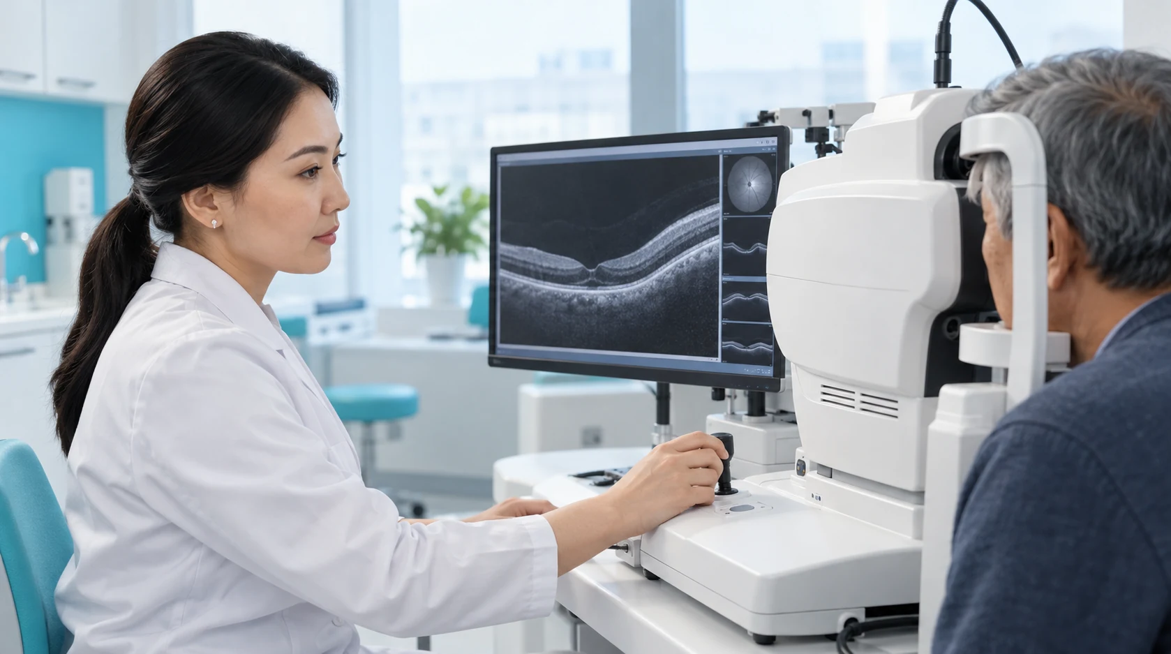
The pupil is usually dilated and topical anaesthetic drops used.

The patient is positioned at the laser; a contact diagnostic lens may be placed on the eye.
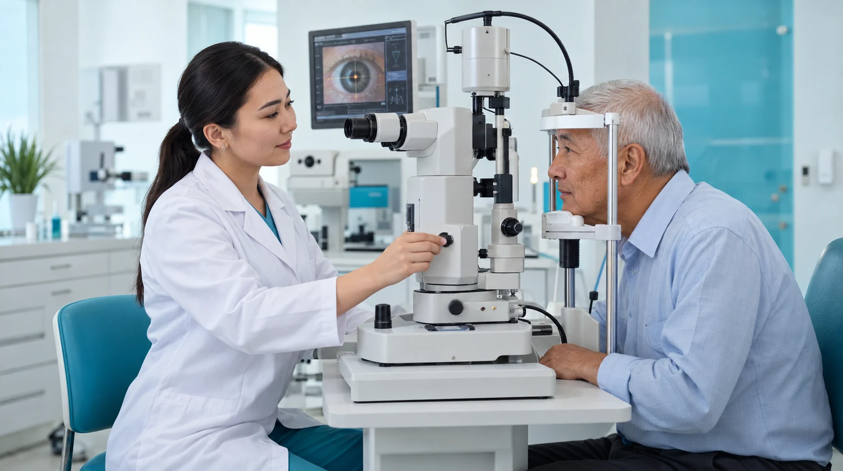
The doctor applies coagulation spots according to the selected pattern.
The doctor assesses the eye, gives advice and schedules review.

Laser treatment is directed at specific retinal areas according to the diagnosis and examination results.
Standard retinal laser photocoagulation is performed without surgically opening the eye.
In complex disease, laser treatment can complement other indicated retinal treatments.
The extent and placement of laser spots are selected according to the type of retinal damage.
Laser photocoagulation creates controlled treatment spots in selected retinal areas.
Learn moreBook a consultation with a KazRIED laser ophthalmic surgeon. The doctor will examine the retina, determine whether laser is indicated, select the appropriate technique and decide whether treatment should be completed in one or several stages.
A team of experienced doctors across specialties.

Candidate of Medical Sciences; Highest-category ophthalmologist
Highest-category ophthalmologist
Highest-category ophthalmologist
Highest-category ophthalmologist

Second-category ophthalmologist

PhD Student


Laser photocoagulation creates controlled treatment spots in selected areas of the retina. Laser energy produces a localized thermal reaction. In the peripheral retina, this helps form chorioretinal adhesion around an area at risk.
The goal depends on the diagnosis: creating a barrier around a tear, reducing the stimulus for abnormal vessel growth, or treating a localized area of disease.
Laser is not a universal way to “strengthen the retina.” Not every area of thinning, case of high myopia, or pregnancy requires photocoagulation.
For significant retinal detachment, outpatient laser photocoagulation usually does not replace vitreoretinal surgery.
Laser may also be applied inside the eye during surgery as endolaser treatment. This is a different clinical setting from outpatient photocoagulation.
The decision follows a fundus examination and any necessary diagnostic tests. Careful peripheral retinal examination is important; vascular disease also requires assessment of the macula, ischemia, and disease activity.
The clinician applies laser spots according to the selected pattern. The number of pulses and treatment area depend on the diagnosis.
Afterward, the eye is assessed, instructions are provided, and follow-up is arranged. Panretinal treatment often requires several sessions.
Laser spots create a local barrier but do not prevent new tears elsewhere in the retina.
Recurrent flashes, a new curtain-like shadow, or a sudden increase in floaters require urgent reassessment, even if previous treatment was successful.
In diabetic retinopathy, treatment often forms part of long-term monitoring. Managing blood glucose, blood pressure, and other systemic factors affects progression risk but does not replace ophthalmic treatment.
PRP, anti-VEGF therapy, and surgery serve different purposes. The treatment plan may change as the retinal condition changes.
Kazakhstan’s first specialized ophthalmology institute, operating since 1933.
Modern diagnostics and surgery using world-class equipment.
Leading ophthalmologists in Kazakhstan, scientists and experienced surgeons.
More than 30,000 operations and around 270,000 consultations annually.


Kazakhstan’s first specialized ophthalmology institute, operating since 1933.
Modern diagnostics and surgery using world-class equipment.
Leading ophthalmologists in Kazakhstan, scientists and experienced surgeons.
More than 30,000 operations and around 270,000 consultations annually.
Kazakhstan’s first specialized ophthalmology institute, operating since 1933.
Modern diagnostics and surgery using world-class equipment.
Leading ophthalmologists in Kazakhstan, scientists and experienced surgeons.
More than 30,000 operations and around 270,000 consultations annually.


