


Vision has become cloudy
A veil, fog or gradually reduced clarity may be associated with cataract, but similar symptoms occur in other diseases, so an examination is needed.
Cataract treatment replaces the cloudy natural lens with an artificial lens. The operation and IOL are selected after examination and discussion of expected vision.
A veil, fog or gradually reduced clarity may be associated with cataract, but similar symptoms occur in other diseases, so an examination is needed.
Oncoming headlights, lamps or sunlight may cause marked discomfort and reduce visual quality.
Frequent prescription changes or inability to regain previous clarity with glasses are reasons to examine the lens and other eye structures.
If optical correction no longer provides acceptable visual function, the doctor assesses the benefit of surgery.
Dense clouding can make retinal examination or necessary treatment of other eye conditions difficult.
Macular degeneration, diabetic retinopathy and other retinal diseases can limit final visual acuity.
Lens replacement removes the cataract but does not restore damage to the optic nerve or retina.
Corneal curvature and transparency affect IOL calculations and visual quality after surgery.
Laser vision correction, vitreoretinal surgery and other procedures may alter the calculations and surgical plan.
Retinal health and systemic disease control need assessment because diabetic disease can affect the prognosis and postoperative follow-up.
Cataract surgery is sometimes combined with glaucoma surgery or requires separate intraocular-pressure monitoring.
Additional fixation, a capsular tension ring or another surgical approach may be needed.
The surgeon assesses complexity and additional risks in advance to select a safe technique and consumables.
Comprehensive diagnostics are needed before surgery. Cataract may be the main cause of reduced vision, but retinal, optic-nerve or corneal disease may coexist. In such cases, cataract removal eliminates the cloudy lens but does not guarantee the visual acuity the patient expects.
Reduced contrast and night vision can significantly affect driving and orientation in low light.
Surgery is considered when reduced vision makes reading, work, driving, mobility, household tasks or other important activities difficult.
In some situations, cataract can be associated with raised intraocular pressure or other complications that affect the timing and surgical approach.
Preparing for cataract surgery
As with any intraocular operation, complications are possible.
The doctor must discuss individual risks, the possible need for glasses after surgery, the features of the selected IOL and symptoms that require urgent medical attention after treatment.
As with any intraocular operation, complications are possible. The doctor must discuss individual risks, the possible need for glasses after surgery, the features of the selected IOL and symptoms that require urgent medical attention after treatment.
Book a consultation
The doctor confirms the cataract and assesses visual acuity and quality, intraocular pressure, and the anterior and posterior segments.
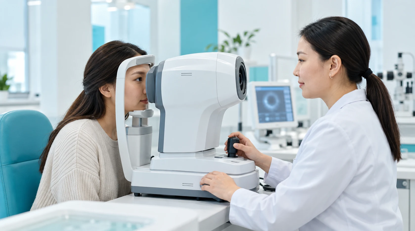
Eye length and corneal parameters are measured and IOL power is calculated.

The patient receives a list of tests and instructions on medications, food and preparation on the day of surgery.

The opaque lens is fragmented and removed through a small surgical opening.
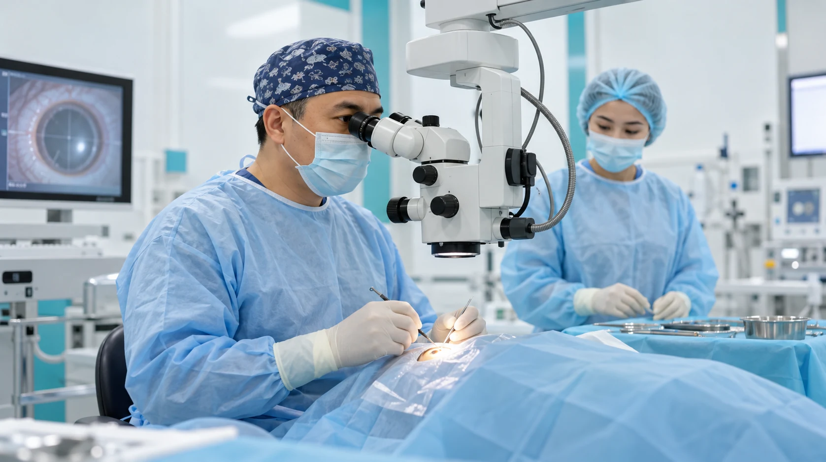
After surgery, the doctor assesses IOL position and the eye and prescribes drops and temporary restrictions.

Removing the cloudy lens can improve vision within the capabilities of the retina and optic nerve.
After the cloudy lens is treated, objects may appear clearer and colours easier to distinguish.
The artificial lens is selected according to eye health and the patient's visual needs.
Diagnostic assessment helps plan surgery and estimate the possible outcome when other eye conditions are present.
A monofocal IOL has one principal optical focus.
Learn moreA toric IOL replaces the lens while correcting corneal astigmatism.
Learn moreA multifocal IOL provides several optical foci to reduce dependence on glasses at different distances.
Learn moreFind out whether you need cataract surgery Start with diagnostics. The doctor will determine whether cataract is actually causing the visual loss, assess the retina and optic nerve, and explain which treatment and IOL options may be considered for you.
A team of experienced doctors across specialties.

Doctor of Medical Sciences, Associate Professor; Highest-category ophthalmologist
Candidate of Medical Sciences, Associate Professor; Highest-category ophthalmologist

Candidate of Medical Sciences; Highest-category ophthalmologist

Candidate of Medical Sciences; Highest-category ophthalmologist

Candidate of Medical Sciences; Highest-category ophthalmologist

Candidate of Medical Sciences; Highest-category ophthalmologist

Doctor of Philosophy (PhD)
Master’s Student in Public Health

Highest-category ophthalmologist

Highest-category ophthalmologist
A cataract is a clouding of the crystalline lens, the transparent natural lens inside the eye. The clouded lens transmits and focuses light less effectively, making images blurred and gradually impairing vision.
Cataracts most commonly develop with age. They can also result from eye injury, diabetes, prolonged use of certain medications, or inflammatory eye diseases. Cataracts may also be congenital.
Medications and eye drops cannot restore transparency to an already clouded lens. The only way to remove a cataract is to surgically replace the natural lens with an artificial intraocular lens (IOL).
The main modern method is phacoemulsification: the clouded lens is removed through a small surgical incision and replaced with an IOL.
If a cataract interferes with reading, driving, work, or other everyday activities, the clinician may recommend surgery.
There is no need to wait for the cataract to become “ripe.” The decision considers its impact on vision and daily life, along with the risks and expected benefits for the individual patient.
Before surgery, KazRIED performs a diagnostic assessment and calculates the required IOL power.
The IOL type is selected according to the eye’s anatomy, astigmatism, the condition of the retina and optic nerve, the patient’s usual visual demands, and their expectations.
Assessment confirms whether visual loss is caused by clouding of the lens, evaluates other eye structures and provides information for surgical planning.
The main surgical method for cataract removal: the cloudy lens is fragmented and removed, then an IOL is implanted.
A separate pathway for patients whose operation requires additional planning because of dense cataract, weak lens-supporting ligaments, coexisting eye disease or other risk factors.
Clouding of the posterior capsule after previous surgery can reduce vision again. After examination, the doctor may recommend YAG laser capsulotomy.
IOL selection affects the optical target of surgery, the need for glasses after treatment and the possibility of correcting astigmatism.
The doctor confirms the cataract and assesses visual acuity and quality, intraocular pressure, and the anterior and posterior segments. OCT, ultrasound and other examinations may be ordered to establish what limits vision and what outcome can be expected.
Eye length and corneal parameters are measured and IOL power is calculated. The surgeon discusses the planned main focal distance and whether a toric, EDOF or multifocal option is needed.
The patient receives a list of tests and instructions on medications, food and preparation on the day of surgery. Coexisting diseases and medications must be discussed with the doctor.
The opaque crystalline lens is fragmented and removed through a small surgical opening. A calculated IOL is implanted into the retained capsule where possible. Technique depends on the eye's condition.
After surgery, the doctor assesses IOL position and the eye and prescribes drops and temporary restrictions. Reviews track healing and detect complications promptly.
Biometry and keratometry are used to calculate the optical power of the artificial lens and plan the refractive outcome.
The surgeon and, when necessary, an anesthesiologist review test results, coexisting medical conditions, and regularly taken medications.
The medication regimen is prescribed individually. Do not use someone else’s eye drops or previously prescribed drops without consulting your clinician.
The timing of treatment for the second eye is discussed separately after assessing the first eye and the patient’s visual needs.
The comparison describes classes of intraocular lenses (IOLs), rather than specific models. Toric optics can be combined with different focal designs and do not represent a separate level of lens quality.
An intraocular lens (IOL) is selected according to the patient’s visual needs and the condition of the eye.
The key question is which optical outcome the individual patient needs and whether the eye’s condition allows the selected lens design to be used safely.
The clinician assesses not only the cataract, but also the cornea, macula, optic nerve, astigmatism, and previous surgeries.
The patient’s usual activities are also considered: reading, using a phone or computer, driving, and tasks involving distance or near vision.
Related information: Astigmatism.
Kazakhstan’s first specialized ophthalmology institute, operating since 1933.
Modern diagnostics and surgery using world-class equipment.
Leading ophthalmologists in Kazakhstan, scientists and experienced surgeons.
More than 30,000 operations and around 270,000 consultations annually.


Kazakhstan’s first specialized ophthalmology institute, operating since 1933.
Modern diagnostics and surgery using world-class equipment.
Leading ophthalmologists in Kazakhstan, scientists and experienced surgeons.
More than 30,000 operations and around 270,000 consultations annually.
Kazakhstan’s first specialized ophthalmology institute, operating since 1933.
Modern diagnostics and surgery using world-class equipment.
Leading ophthalmologists in Kazakhstan, scientists and experienced surgeons.
More than 30,000 operations and around 270,000 consultations annually.


