

Retinal tears and detachment
Rhegmatogenous detachment with one or more retinal breaks. Limited local detachment or tear where laser barricade or external buckle is being considered.
Retinal detachment requires urgent vitreoretinal assessment. Surgery is chosen according to the detachment type and extent, retinal breaks and other eye characteristics.
Rhegmatogenous detachment with one or more retinal breaks. Limited local detachment or tear where laser barricade or external buckle is being considered.
Tractional detachment, including proliferative diabetic retinopathy.
Combined tractional-rhegmatogenous detachment.
Recurrent detachment after previous surgery.
Detachment with vitreous haemorrhage, PVR, multiple breaks or macular involvement.
Exudative detachment requiring investigation and treatment of the underlying cause.
Retinal detachment is not treated with eye drops, vitamins or vascular-support courses. In established rhegmatogenous or tractional detachment, external laser alone does not replace vitreoretinal surgery. Even after successful anatomic reattachment, visual recovery may be incomplete, particularly after prolonged macular detachment, ischaemia or advanced PVR.
Sudden flashes, a marked increase in floaters, a dark shadow or curtain, field loss or rapid visual decline require urgent dilated retinal examination rather than waiting for a routine visit. Bring records of previous eye surgery, IOLs, silicone oil or prior retinal tears. Do not stop anticoagulants, antiplatelets or systemic medicines on your own.
Retinal detachment is not treated with eye drops, vitamins or vascular-support courses. In established rhegmatogenous or tractional detachment, external laser alone does not replace vitreoretinal surgery. Even after successful anatomic reattachment, visual recovery may be incomplete, particularly after prolonged macular detachment, ischaemia or advanced PVR. With intraocular gas, air travel and major altitude exposure are prohibited until the gas is fully absorbed; any anaesthetist must be told about the gas bubble.
Book a consultation
Visual acuity, pressure, biomicroscopy and dilated fundus examination.

Tear location, detachment extent, macular status, PVR and vitreous; ultrasound for opaque media and macular OCT when informative.

Laser, external buckling, vitrectomy or a combination; discussion of tamponade and anaesthesia.

Preoperative preparation under the KazRIED protocol and assessment of systemic risk.

Closing the tear or releasing traction, flattening the retina, retinopexy and gas, silicone or perfluorocarbon liquid tamponade when needed.
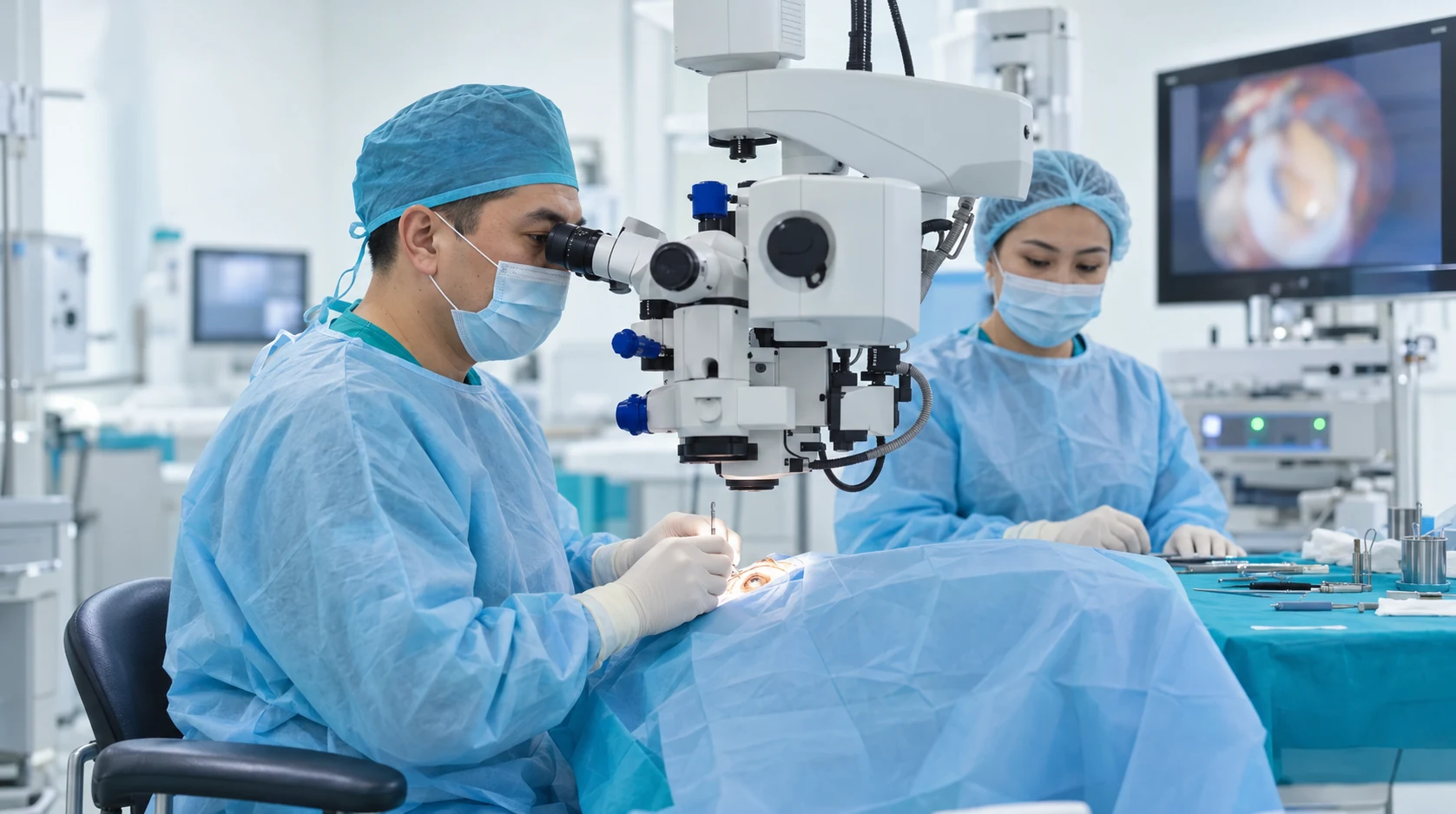
Retinal position, pressure, inflammation, macular and tamponade status.

Monitoring recurrence, new tears and the fellow eye; separate silicone removal when required.
Surgery aims to return the detached retina to its correct anatomical position.
Timely treatment aims to preserve available visual function and reduce the risk of further deterioration.
Vitreoretinal surgery makes it possible to treat changes that cannot be addressed with drops or external laser alone.
Follow-up examinations help assess the retinal response to treatment and identify the need for further stages in time.
Colour applies only to the individual criterion: green = more favourable; yellow = situation-dependent; red = limitation; grey = neutral/not directly comparable. It is not an overall ranking.
Colour applies only to the individual criterion: green = more favourable; yellow = situation-dependent; red = limitation; grey = neutral/not directly comparable. It is not an overall ranking.
If retinal detachment is suspected, arrange an urgent KazRIED vitreoretinal surgeon examination. After imaging and retinal examination, the surgeon determines urgency and whether laser, scleral indentation, vitrectomy or combined surgery is appropriate.
A team of experienced doctors across specialties.

Doctor of Medical Sciences; Highest-category ophthalmologist
Candidate of Medical Sciences, Associate Professor; Highest-category ophthalmologist

Doctor of Philosophy (PhD)

Highest-category ophthalmologist

Highest-category ophthalmologist

First-category ophthalmologist

First-category ophthalmologist

Second-category ophthalmologist; PhD Degree Candidate


Retinal detachment is the separation of the neurosensory retina from the underlying retinal pigment epithelium. Treatment depends on the type of detachment and the condition of the individual eye.
Treatment planning considers:
KazRIED uses vitrectomy, scleral buckling, and laser methods in selected, limited situations.
The goal is to restore anatomical retinal attachment, address the cause of detachment, and preserve as much visual function as possible. In rhegmatogenous detachment, the source of fluid entering beneath the retina must be sealed.
Urgency depends primarily on the risk of macular involvement and the duration of detachment.
Patients should not try to judge the stage by symptom severity: a peripheral detachment can progress rapidly, while symptoms of tractional or exudative detachment may develop gradually.
After surgery, monitoring includes retinal attachment, intraocular pressure, macular condition, and the tamponade agent.
If gas is used, the clinician may prescribe a specific head position and prohibit air travel. Silicone oil may need to be removed in a separate procedure after the retina has stabilized.
The timing of return to work, driving, and physical activity is determined individually.
Kazakhstan’s first specialized ophthalmology institute, operating since 1933.
Modern diagnostics and surgery using world-class equipment.
Leading ophthalmologists in Kazakhstan, scientists and experienced surgeons.
More than 30,000 operations and around 270,000 consultations annually.


Kazakhstan’s first specialized ophthalmology institute, operating since 1933.
Modern diagnostics and surgery using world-class equipment.
Leading ophthalmologists in Kazakhstan, scientists and experienced surgeons.
More than 30,000 operations and around 270,000 consultations annually.
Kazakhstan’s first specialized ophthalmology institute, operating since 1933.
Modern diagnostics and surgery using world-class equipment.
Leading ophthalmologists in Kazakhstan, scientists and experienced surgeons.
More than 30,000 operations and around 270,000 consultations annually.


