Almaty, Astana, Shymkent

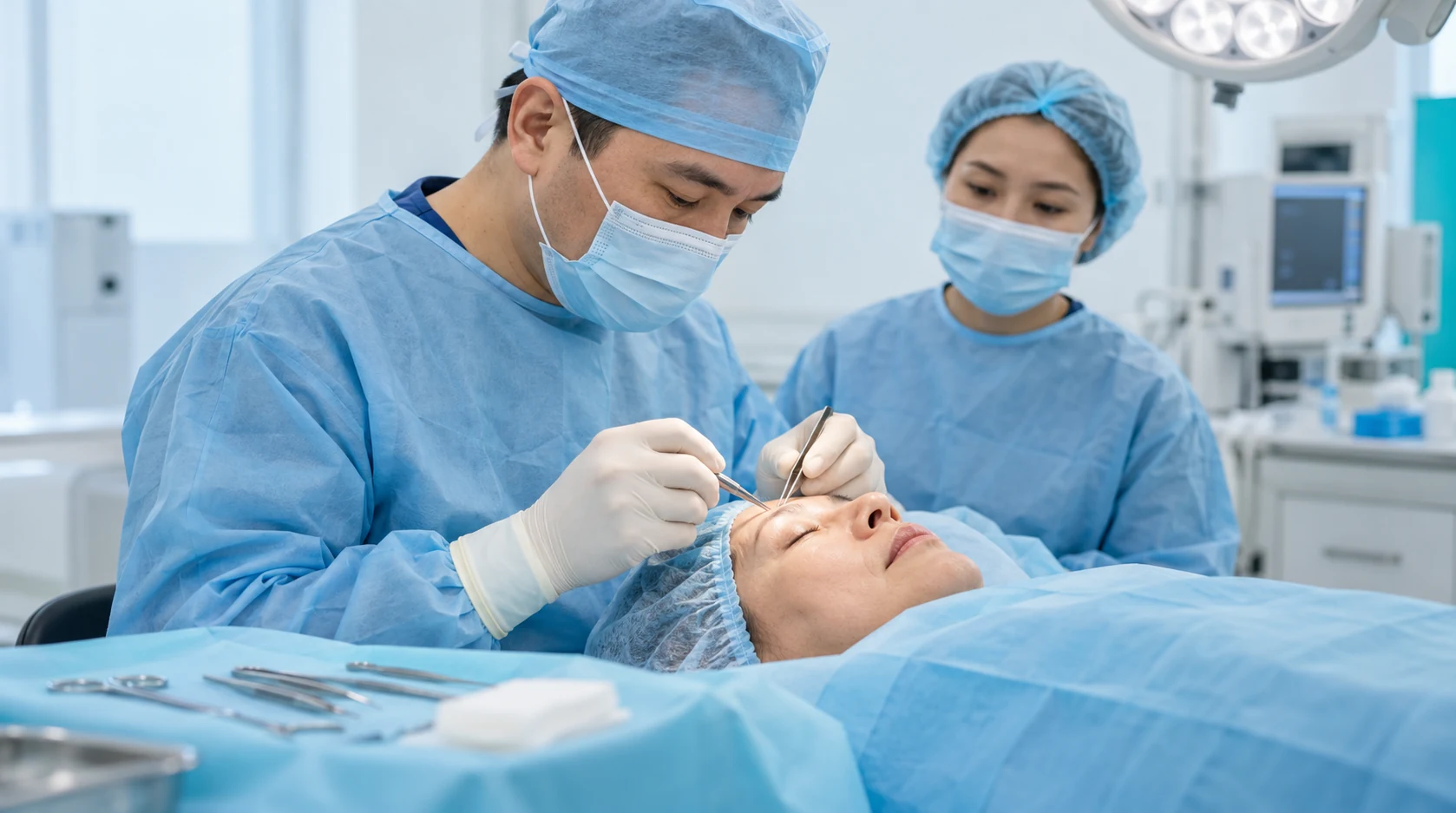
Eyelid Lesion Removal

from 45.500 ₸
Book an appointment

About the service

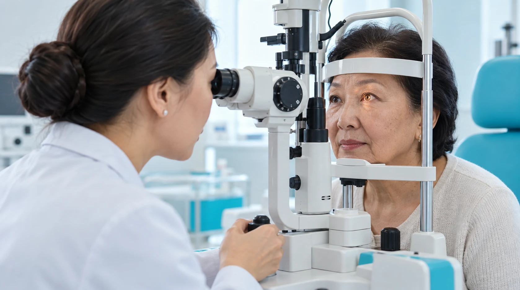
For an eyelid lesion, the ophthalmologist assesses its features and chooses monitoring, biopsy or removal. The procedure depends on location, suspected diagnosis and the need for tissue examination.

Who the service is for

Eye injury

a benign lesion that is repeatedly traumatized, irritates the ocular surface, interferes with blinking or causes meaningful cosmetic concern; In the eyelid area, there is a risk of eye injury and of destroying tissue that may be needed for histopathologic diagnosis. fixation or spread into surrounding tissue;

Changes in an eyelid lesion

growth of the lesion or changes in its surface, shape or pigmentation; rapid growth; change in color or shape; ulceration or a non-healing surface; spontaneous bleeding; recurrence after removal;

Optical correction

bleeding, ulceration or persistent crusting without a clear benign explanation;

Eyelid condition

loss of eyelashes or distortion of the normal eyelid margin; Arrange an examination if a new nodule, plaque, pigmented spot, growth or gradually enlarging lesion appears on the upper or lower eyelid. distortion of the eyelid margin;

Previous treatment

recurrence after previous removal;

Eyelid lesion

an uncertain diagnosis requiring biopsy and histopathology; a suspected malignant or premalignant lesion requiring a pathology-driven treatment pathway.

Eyelid condition

Arrange a more prompt examination for accelerated growth, changes in colour or shape, ulceration, bleeding, lash loss, distortion of the eyelid contour, recurrence or hardening of surrounding tissues.

Reduced vision

new visual loss, double vision, severe pain or restricted eye movement.

Limitations and safety

Қабақ ісігін жоспарлы түрде алып тастау операциясы көздің немесе оған жанасатын терінің өтіп жатқан жіті инфекциясы, жалпы жіті аурулар немесе операциялық қауіпті арттыратын декомпенсацияланған жағдайлар кезінде шегерілуі мүмкін. Қанның ұюына әсер ететін препараттарды қабылдауды алдын ала дәрігермен талқылау қажет. Салыстырмалы қарсы көрсеткіштер алып тастау әдісін, ақаудың көлемін, реконструкция қажеттілігін, анестезияны және пациенттің жалпы жағдайын ескере отырып, жеке анықталады. Түзілістің жылдам өсуі, түсінің немесе пішінінің өзгеруі, жазылмайтын ойық жара, өздігінен қан кету, кірпіктердің түсуі, қабақтың деформациясы, тығыз бекітілуі, айналадағы тіндерге таралуы немесе қайталануы (рецидив) шұғыл тексеруді талап етеді. Көру қабілетінің жаңадан төмендеуі, екіге көріну, қатты ауырсыну немесе көз қозғалысының шектелуі байқалса, дереу медициналық көмекке жүгіну қажет.

Book a consultation

Stages

Stage 1

Consultation

The ophthalmologist or ophthalmic surgeon reviews the lesion history and previous procedures.

Consultation

Service benefits

Informed lesion removal

The doctor assesses the lesion and selects a removal method according to its location and suspected nature.

An opportunity to clarify the lesion's nature

When indicated, removed tissue is sent for histological examination to clarify the diagnosis.

Protecting the ocular surface

Treatment aims to reduce corneal irritation and restore eyelid protection according to the cause of the problem.

Consideration of eyelid function

Removal and, when needed, reconstruction are planned to preserve eye closure and protection.

Methods comparison

Criterion
Surgical excision / excisional biopsy
Incisional biopsy
Cryodestruction
Ablative method without an intact specimen
Primary purpose
Remove a localized lesion and obtain tissue
Establish diagnosis before definitive treatment
Destroy selected tissue by freezing
Remove or destroy tissue locally
Tissue for histology
Usually provides an intact specimen
Yes, this is the main diagnostic purpose
An intact specimen is generally not obtained
May be limited or unavailable
Suspicion of malignancy
May be appropriate within an oncologic surgical plan
Commonly used for diagnostic confirmation
Should not be chosen simply for cosmetic removal without diagnosis
Not a universal choice for a suspicious lesion
Size and anatomy
Depends on ability to close the defect and preserve lid function
Useful when complete removal should not be the first step
Selected indications only
Depends on tissue type and location
Eyelid reconstruction
May be required for a substantial defect
Usually does not provide definitive reconstruction
Does not create the same excisional defect, but tissue changes may occur
Depends on treatment depth
Margin assessment
Can be integrated into surgical and pathology planning
Not definitive removal
Histologic margin control is limited
Histologic margin control may be impossible
Chance of another procedure
Depends on diagnosis and excision margins
A definitive treatment step commonly follows pathology
Another procedure may be needed if tissue destruction is incomplete or the indication was selected incorrectly.
Depends on diagnosis and depth
Scar / lid contour change
Possible, especially with larger defects; reconstruction may be needed
Smaller tissue sample, but a biopsy site remains
Pigment change, scarring or local contour change can occur
Risk depends on method and depth
Recovery
From several days to a longer period, depending on the extent of the procedure and reconstruction.
Often smaller procedure, but treatment is not yet complete
Depends on depth and tissue response
No universal recovery time
Published KazRIED price
No universal tariff; selected lesion-specific tariffs are published
No separate public tariff found under this name
from 32 500 ₸
Exact method and tariff require clinic confirmation
Surgical excision / excisional biopsy
Primary purpose
Tissue for histology
Suspicion of malignancy
Size and anatomy
Eyelid reconstruction
Margin assessment
Chance of another procedure
Scar / lid contour change
Recovery
Published KazRIED price
Remove a localized lesion and obtain tissue
Usually provides an intact specimen
May be appropriate within an oncologic surgical plan
Depends on ability to close the defect and preserve lid function
May be required for a substantial defect
Can be integrated into surgical and pathology planning
Depends on diagnosis and excision margins
Possible, especially with larger defects; reconstruction may be needed
From several days to a longer period, depending on the extent of the procedure and reconstruction.
No universal tariff; selected lesion-specific tariffs are published
Establish diagnosis before definitive treatment
Yes, this is the main diagnostic purpose
Commonly used for diagnostic confirmation
Useful when complete removal should not be the first step
Usually does not provide definitive reconstruction
Not definitive removal
A definitive treatment step commonly follows pathology
Smaller tissue sample, but a biopsy site remains
Often smaller procedure, but treatment is not yet complete
No separate public tariff found under this name
Destroy selected tissue by freezing
An intact specimen is generally not obtained
Should not be chosen simply for cosmetic removal without diagnosis
Selected indications only
Does not create the same excisional defect, but tissue changes may occur
Histologic margin control is limited
Another procedure may be needed if tissue destruction is incomplete or the indication was selected incorrectly.
Pigment change, scarring or local contour change can occur
Depends on depth and tissue response
from 32 500 ₸
Remove or destroy tissue locally
May be limited or unavailable
Not a universal choice for a suspicious lesion
Depends on tissue type and location
Depends on treatment depth
Histologic margin control may be impossible
Depends on diagnosis and depth
Risk depends on method and depth
No universal recovery time
Exact method and tariff require clinic confirmation

Book an appointment

Book a consultation with a KazRIED ophthalmic surgeon. The doctor will assess the lesion, determine whether biopsy or removal is needed and explain which method is suitable for your situation.

Book an appointment

Procedure prices

Service name Price
Eyelid papilloma removal (per eye) from 45 500 ₸
Xanthelasma removal (per eye) from 45 500 ₸
Removal of atheroma, pinguecula and molluscum contagiosum (per eye) from 45 500 ₸
Cryodestruction (per eye) from 32 500 ₸
Histologic examination of surgical material, including the eyeball (per eye) from 21 000 ₸
Cytologic examination (per eye) from 5 200 ₸
Our services, payable in installmentsTake care of your vision today and pay for our services in installments. Choose installment terms and a payment schedule that suit you.

Our doctors

A team of experienced doctors across specialties.

All doctorsThe full list of doctors is available here!

Eyelid lesions: diagnosis, removal, and tissue examination

Similar-looking eyelid lumps and growths can have different origins. The eyelid consists of skin, muscle, a tarsal plate containing glands, and conjunctiva. Lesions can arise from different tissues and may be benign, precancerous, or malignant.

What is assessed at the first appointment?

The main goal is to establish the clinical nature of the lesion. The clinician assesses its duration, growth, color, surface, eyelid margin and eyelashes, attachment to surrounding tissues, and other features.

If malignancy is suspected, pathological confirmation and an oncologically appropriate treatment approach are required. The removal method must preserve the opportunity for tissue examination.

When is complete excision appropriate, and when is a biopsy needed?

A small lesion suitable for complete excision can sometimes be removed in one procedure. This can serve both diagnostic and therapeutic purposes.

For a large, infiltrative, diagnostically uncertain, or complexly located lesion, the clinician may perform a biopsy first. This clarifies the diagnosis and helps plan excision and reconstruction without unnecessarily compromising eyelid anatomy.

Laser treatment, radiofrequency treatment, and cryodestruction are not universal methods for all lesions.

How does this service differ from chalazion removal?

A chalazion is an inflammatory condition of a meibomian gland. KazRIED lists its removal under a separate tariff.

A recurrent or atypical lesion resembling a chalazion may require additional assessment and tissue examination.

Assessment, preparation, and anesthesia

The clinician examines the eyelid and surrounding structures. Additional tests are requested when clinically indicated.

Required tests and medication restrictions depend on the extent of surgery and the type of anesthesia. Do not stop anticoagulants or other medications without medical advice.

Local anesthesia is often used for small procedures. The clinician determines the specific approach; anesthesia planning may differ for children and complex operations.

Which circumstances affect treatment planning?

  • A newly developed lump or growth: the diagnosis and need for removal must be established.
  • Recurrence after removal: previous histology findings and the cause of recurrence are important.
  • Previous eyelid surgery: scarring and altered anatomy may affect surgical access, excision extent, and reconstruction.
  • A child: lesion type, visual impact, and anesthesia requirements are considered jointly by a pediatric ophthalmologist and surgeon.
  • An older patient: particular attention is paid to medications, coexisting conditions, and possible signs of malignancy.
  • Travel from another city: clarify whether consultation, surgery, and histological examination can be arranged within one trip or require several stages.

Examination of removed tissue

Removed tissue is submitted for pathological examination according to the treatment plan. KazRIED has its own pathology laboratory.

Histological examination of surgical specimens is a separate laboratory service. The tariff item also mentions specimens from the eyeball; this does not mean that removing an eyelid lesion involves removing the eye.

Cytological examination is a separate laboratory service and is not required in every case.

What happens after the results are available?

If the lesion is benign and there are no concerns about excision margins, subsequent follow-up is usually more straightforward.

If a malignant tumor or precancerous change is found, further excision, reconstruction, lymph node assessment, or shared care with an oncologist may be needed.

Consult an ophthalmic surgeon before cosmetic procedures around the eyelid, particularly if a lesion is growing, bleeding, changing pigmentation, or disrupting the eyelash margin.

Facts and offers

Kazakhstan’s first specialized ophthalmology institute, operating since 1933.

Modern diagnostics and surgery using world-class equipment.

Leading ophthalmologists in Kazakhstan, scientists and experienced surgeons.

More than 30,000 operations and around 270,000 consultations annually.

Related services

Frequently asked questions

Is eyelid lesion removal painful?
Anaesthesia is used before the procedure. Small operations in adults are often performed under local anaesthesia. The anaesthetic injection may briefly be uncomfortable, after which pain sensation in the operative area is substantially reduced.
What type of anaesthesia is used?
It depends on lesion size and location, surgical extent, need for reconstruction, age and general health. Local anaesthesia, local anaesthesia with sedation or general anaesthesia may be used in selected situations.
How long does eyelid lesion removal take?
There is no universal duration. It depends on lesion size and depth, eyelid-margin involvement, the type of biopsy or excision, need for reconstruction and anaesthesia.
Can I go home on the day of surgery?
This is often possible after a minor procedure. The decision depends on anaesthesia, surgical extent, reconstruction, general health and early postoperative recovery.
How should I prepare for eyelid lesion removal?
Assessment and diagnosis come first. Tell the clinician about all medicines, allergies and medical conditions. Eye-area makeup is generally avoided on the procedure day. Additional preparation depends on the surgery and anaesthesia.

Confirm you are not a robot

Complete the check to send your request.