

Visual field
Patients with a symptomatic retinal tear or another peripheral lesion judged high risk by a retina specialist. Patients with selected local peripheral detachment that can be safely demarcated with laser.
Laser photocoagulation creates controlled treatment spots in selected retinal areas. Its purpose and pattern depend on the condition, from surrounding high-risk changes to treating ischaemic and vascular disease.
Patients with a symptomatic retinal tear or another peripheral lesion judged high risk by a retina specialist. Patients with selected local peripheral detachment that can be safely demarcated with laser.
Patients with preproliferative or proliferative diabetic retinopathy when panretinal photocoagulation is indicated.
Patients with ischaemic changes after retinal vein occlusion when laser is part of the treatment plan.
Patients with selected local vascular or macular lesions where focal laser has a defined clinical purpose.
Patients after previous retinal laser when follow-up shows new areas requiring additional treatment.
Not every peripheral degeneration or asymptomatic lesion requires prophylactic laser. High myopia or pregnancy alone is not a sufficient indication without a retinal lesion that warrants treatment. Established extensive retinal detachment usually cannot be managed by laser alone. Large-volume panretinal photocoagulation can affect peripheral and night vision;
Large-volume panretinal photocoagulation can affect peripheral and night vision; temporary blur from dilation, inflammation, retinal oedema or IOP change can occur after laser.
The pupil may be dilated on the treatment day, so confirm driving and accompaniment arrangements in advance.
Seek urgent review for severe pain, sudden visual loss, a new curtain or a marked increase in flashes and floaters. If you develop new flashes, a sudden shower of floaters, a dark curtain or a new visual-field defect, you need urgent retinal assessment rather than a routine laser booking.
Bring previous OCT/OCT-A, fluorescein angiography, ultrasound and records of retinal laser or vitreoretinal surgery.
If you have diabetes, recent glucose and HbA1c data are useful.
Not every peripheral degeneration or asymptomatic lesion requires prophylactic laser.
Book a consultation
Symptoms, duration, diabetes, vascular disease and previous laser or surgical treatment.

Fundus examination with a special contact lens and a dilated pupil; OCT, OCT angiography, fluorescein angiography, ultrasound or other tests are added when necessary.
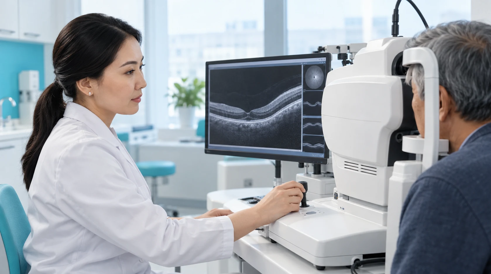
Prophylactic peripheral coagulation or barrage, panretinal, focal, paravascular, sectoral or another indicated method.

On the day of the procedure, the pupil is dilated, local anaesthetic drops are used and a special lens is placed on the eye.

The number of burns and treatment stages is not fixed universally.
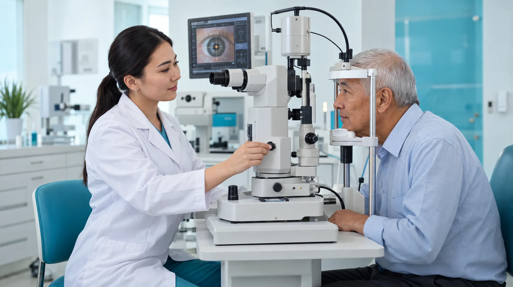
A check is performed after the procedure; the timing of the next examination and restrictions depend on the diagnosis, extent of photocoagulation and coexisting conditions.

For suitable peripheral tears, laser helps form adhesions around the area and reduce the risk of detachment spreading.
In suitable retinopathies, laser photocoagulation helps control the effects of ischaemia and abnormal blood vessel growth.
Laser treatment is directed at specific retinal areas according to the diagnosis and examination results.
Follow-up examinations help assess the retinal response to treatment and identify the need for further stages in time.
Colour applies only to the individual criterion: green = more favourable; yellow = situation dependent; red = limitation; grey = neutral/not directly comparable. It is not an overall ranking.
Colour applies only to the individual criterion: green = more favourable; yellow = situation dependent; red = limitation; grey = neutral/not directly comparable. It is not an overall ranking.
Book a consultation with a KazRIED retinal laser surgeon. After examining the retina, the doctor will decide whether laser is indicated, which pattern is appropriate and whether one session, staged treatment or another method is required.
A team of experienced doctors across specialties.

Candidate of Medical Sciences; Highest-category ophthalmologist
Highest-category ophthalmologist
Highest-category ophthalmologist

PhD Student

Laser photocoagulation creates localized thermal burns in selected areas of the retina. The type and extent of treatment depend on the diagnosis.
For retinal tears and selected peripheral lesions, laser burns are placed around the area at risk to form chorioretinal adhesions and limit the spread of fluid beneath the retina.
In preproliferative and proliferative diabetic retinopathy, panretinal photocoagulation is used when indicated to treat ischemic retinal areas and reduce the stimulus for pathological neovascularization.
Focal, paravascular, or sectoral photocoagulation is used for more localized treatment goals.
Laser treatment does not restore retinal cells that have already died. It does not replace vitrectomy for extensive retinal detachment, marked traction, or opaque media that prevent safe visualization of the target.
Retinal photocoagulation is not intended to change the glasses prescription and does not replace corneal laser vision correction.
The laser surgeon determines the type and extent of treatment after a dilated fundus examination and additional tests when indicated.
The treatment goal depends on the retinal findings: creating a preventive barrier, treating an ischemic area, or performing localized photocoagulation for a specific diagnosis.
If prophylactic peripheral laser photocoagulation has been recommended before refractive surgery, pregnancy, or another intervention, the final decision should be based on the peripheral retinal findings. The degree of myopia or the planned procedure alone should not be the sole indication.
The monitoring schedule depends on the reason for treatment and its extent. The clinician assesses the laser-treated areas, peripheral retina, macula, and vascular changes.
In diabetic retinopathy, laser treatment does not replace blood glucose and blood pressure management or regular follow-up. Intravitreal medications may be needed when macular edema is also present.
New tears or areas of degeneration may develop outside the previously treated area, so further examinations remain necessary.
Kazakhstan’s first specialized ophthalmology institute, operating since 1933.
Modern diagnostics and surgery using world-class equipment.
Leading ophthalmologists in Kazakhstan, scientists and experienced surgeons.
More than 30,000 operations and around 270,000 consultations annually.


Kazakhstan’s first specialized ophthalmology institute, operating since 1933.
Modern diagnostics and surgery using world-class equipment.
Leading ophthalmologists in Kazakhstan, scientists and experienced surgeons.
More than 30,000 operations and around 270,000 consultations annually.
Kazakhstan’s first specialized ophthalmology institute, operating since 1933.
Modern diagnostics and surgery using world-class equipment.
Leading ophthalmologists in Kazakhstan, scientists and experienced surgeons.
More than 30,000 operations and around 270,000 consultations annually.


