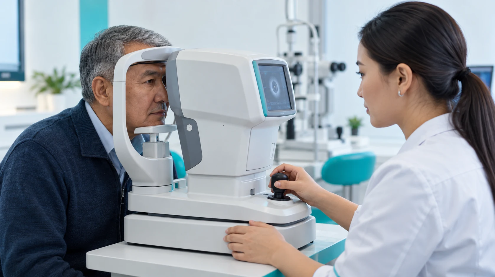
Tonometry measures intraocular pressure using a contact or non-contact method. The result informs eye assessment but does not by itself confirm or exclude glaucoma.
Who the service is for
Intraocular pressure
Patients having a routine eye examination; those with suspected glaucoma or ocular hypertension; people with established glaucoma who need IOP monitoring; and selected patients after ophthalmic procedures as advised by a clinician.
Lens wear schedule
Tonometry may form part of assessment for eye pain, halos, blurred vision, marked redness or other complaints. These symptoms are not specific to high IOP, and acute symptoms require medical assessment.
Symptoms and history
Glaucoma, glaucoma suspect status, ocular hypertension, family history of glaucoma, corticosteroid exposure and selected postoperative states. The clinician determines the monitoring interval.
Corneal thickness
Corneal thickness and biomechanics, refractive or corneal surgery, the specific tonometer, measurement technique and diurnal IOP variation can affect the result. Contact testing may be temporarily unsuitable with epithelial defects or active corneal inflammation. Corneal thickness matters, but should not be used for a simplistic formula-based correction of IOP.
Individual treatment choice
A single high or normal reading does not establish the diagnosis.
Interpreting results
Do not apply one universal “normal range” without considering the method and clinical context. For monitoring, compare results obtained with comparable methods and conditions where possible.
Corneal condition
After LASIK, PRK or other corneal procedures, some methods may underestimate or otherwise alter the reading.
Assessment before choosing treatment
Routine tonometry does not require pupil dilation, although dilation may be needed for other tests during a comprehensive examination.
Contact lenses
No special preparation is usually needed. Remove contact lenses, report medications and previous surgery, relax during the measurement and avoid squeezing the eyelids.
Results and documents
Bring previous intraocular-pressure results, particularly if followed for glaucoma or ocular hypertension. Contact lenses are usually removed before testing; bring a case and glasses.
Glaucoma and eye pressure
Tell the specialist about glaucoma drops, systemic steroids, corneal operations and laser vision correction.
Do not rub the eyes
Do not rub the eyes immediately before testing.
Eye injury
Report any eye injury, severe pain or marked inflammation before the procedure.
Lens wear schedule
Sudden severe eye pain, abrupt vision loss, marked redness, nausea or vomiting with eye symptoms, eye trauma or other acute changes.
When urgent care is needed
A routine tonometry appointment does not replace urgent medical assessment.
Limitations and safety
The tonometer does not diagnose glaucoma automatically.
The specialist reviews the goal, lenses, drops, operations, injuries and factors affecting method selection.
Takes: 1 min
Stage 2
Eye and instrument preparation
Non-contact testing needs no special anaesthesia.
Takes: 1 min
Stage 3
Intraocular-pressure measurement
The patient fixates and avoids squeezing the lids.
Takes: 1 min
Stage 4
Measurement quality check
The specialist checks reproducibility and repeats the measurement if needed.
Takes: 1 min
Stage 5
Explaining the findings
The ophthalmologist compares pressure with other findings to decide on repeat checks, pachymetry, gonioscopy, OCT, perimetry or other tests.
Takes: 2 min
Service benefits
Objective pressure measurement
Tonometry provides an intraocular-pressure reading for diagnosis and follow-up.
Supporting glaucoma detection
Pressure measurements complement optic nerve and visual field assessment when glaucoma is suspected.
Checking treatment effectiveness
Changes in measurements help determine whether treatment is sufficient or needs review.
A method suited to your eye
The doctor selects a suitable tonometry method and considers corneal characteristics when interpreting results.
Method comparison, part 1
Criterion
Non-contact (air-puff)
Goldmann applanation
Maklakov applanation
Rebound tonometry
Dynamic contour
Principle
A brief air pulse deforms the cornea and the device estimates IOP from the corneal response.
A prism applanates a standard corneal area and the required force is measured.
An inked weight applanates the cornea and IOP is derived from the imprint size.
A lightweight probe briefly contacts the cornea; IOP is estimated from rebound/deceleration parameters.
A contour-matched sensor contacts the cornea and records pressure over time.
Corneal contact
No
Yes
Yes
Yes, very brief
Yes
Topical anaesthesia
Usually not required
Usually required
Required
Often not required
Protocol-dependent
Fluorescein / dye
No
Usually fluorescein
Collargol
No
No
Typical role
Rapid screening and routine measurement.
Reference clinical technique in many glaucoma guidelines.
Contact method historically used widely in the region.
Rapid measurement useful in selected outpatient and paediatric settings.
Additional method designed to reduce some corneal-property effects.
Non-contact (air-puff)
Principle
Corneal contact
Topical anaesthesia
Fluorescein / dye
Typical role
A brief air pulse deforms the cornea and the device estimates IOP from the corneal response.
No
Usually not required
No
Rapid screening and routine measurement.
A prism applanates a standard corneal area and the required force is measured.
Yes
Usually required
Usually fluorescein
Reference clinical technique in many glaucoma guidelines.
An inked weight applanates the cornea and IOP is derived from the imprint size.
Yes
Required
Collargol
Contact method historically used widely in the region.
A lightweight probe briefly contacts the cornea; IOP is estimated from rebound/deceleration parameters.
Yes, very brief
Often not required
No
Rapid measurement useful in selected outpatient and paediatric settings.
A contour-matched sensor contacts the cornea and records pressure over time.
Yes
Protocol-dependent
No
Additional method designed to reduce some corneal-property effects.
Method comparison, part 2
Criterion
Non-contact
Goldmann
Maklakov
Rebound
Dynamic contour
Corneal thickness / biomechanics
Must be considered; NCT is affected by corneal properties.
CCT and biomechanics affect the reading; simple correction formulas are not recommended.
Method-specific result affected by corneal properties.
Corneal effects remain, although they differ from GAT.
Some corneal-property effects may be reduced, but the technique is not completely independent of the cornea.
After LASIK / PRK
May underestimate; interpret cautiously.
May underestimate; surgical history matters.
Comparability after refractive surgery is limited.
May be useful as an alternative, but interpretation remains clinical.
Sometimes used as an additional method after corneal procedures.
Diagnoses glaucoma by itself
No
No
No
No
No
Longitudinal monitoring
Yes, if the measurement method is comparable and the doctor understands its limitations.
Yes; widely used in glaucoma protocols.
Yes, when follow-up uses the same method.
Yes, with a consistent protocol.
Yes, with a consistent protocol.
Patient comfort
Brief air puff, no contact.
Brief contact after anaesthesia.
Contact, anaesthesia and imprint measurement.
Very brief contact with a small probe.
Sensor remains in contact during the reading.
KazRIED public price
from 900 ₸
Cost determined after consultation
Cost determined after consultation
Cost determined after consultation
Cost determined after consultation
Practical selection
Useful for rapid initial measurement; unusual readings may require confirmation.
Preferred for standard glaucoma assessment where available and used by protocol.
Use depends on local protocol and clinical purpose.
Useful when quick portable measurement is needed.
Niche additional technique.
Non-contact
Corneal thickness / biomechanics
After LASIK / PRK
Diagnoses glaucoma by itself
Longitudinal monitoring
Patient comfort
KazRIED public price
Practical selection
Must be considered; NCT is affected by corneal properties.
May underestimate; interpret cautiously.
No
Yes, if the measurement method is comparable and the doctor understands its limitations.
Brief air puff, no contact.
from 900 ₸
Useful for rapid initial measurement; unusual readings may require confirmation.
CCT and biomechanics affect the reading; simple correction formulas are not recommended.
May underestimate; surgical history matters.
No
Yes; widely used in glaucoma protocols.
Brief contact after anaesthesia.
Cost determined after consultation
Preferred for standard glaucoma assessment where available and used by protocol.
Method-specific result affected by corneal properties.
Comparability after refractive surgery is limited.
No
Yes, when follow-up uses the same method.
Contact, anaesthesia and imprint measurement.
Cost determined after consultation
Use depends on local protocol and clinical purpose.
Corneal effects remain, although they differ from GAT.
May be useful as an alternative, but interpretation remains clinical.
No
Yes, with a consistent protocol.
Very brief contact with a small probe.
Cost determined after consultation
Useful when quick portable measurement is needed.
Some corneal-property effects may be reduced, but the technique is not completely independent of the cornea.
Sometimes used as an additional method after corneal procedures.
No
Yes, with a consistent protocol.
Sensor remains in contact during the reading.
Cost determined after consultation
Niche additional technique.
Book tonometry at KazRIED
Book an intraocular pressure measurement if your clinician has recommended IOP monitoring or you are undergoing an eye examination. If the result is unusual, the ophthalmologist can determine what testing is needed next.
Our services, payable in installmentsTake care of your vision today and pay for our services in installments. Choose installment terms and a payment schedule that suit you.
Intraocular pressure reflects the balance between aqueous humour production and outflow.
Tonometry provides a quantitative IOP measurement, but a single number is not a diagnosis. High IOP does not always mean glaucoma, and glaucoma can develop with IOP values within the statistical normal range.
The reading is therefore interpreted together with optic nerve findings, OCT, visual fields, gonioscopy, pachymetry and other tests as indicated. For follow-up, the measurement method, corneal properties and timing of the test matter.
Further procedure details
Eye and instrument preparation
Non-contact testing requires no special anaesthesia. Contact-method preparation follows the protocol and may include a local anaesthetic.
Intraocular-pressure measurement
The patient fixates without squeezing the lids. The device takes one or more measurements per eye.
Measurement quality check
The specialist checks reproducibility and repeats if needed. An unusual result must be interpreted in its clinical context.
Additional advice and situations
If IOP is within the expected range
The clinician still considers risk factors and optic nerve findings. Normal IOP does not exclude glaucoma.
If IOP is higher than expected
Repeat measurement, corneal thickness assessment, gonioscopy, OCT and visual fields may be needed. Treatment is not based on a single reading alone.
If two methods give different results
The difference may reflect device principles and corneal properties. The clinician selects the method to use as the main reference for future follow-up.
After refractive surgery
Altered corneal biomechanics and preoperative measurements, if available, should be considered.
If glaucoma treatment is already in use
IOP is compared with the individual target set by the treating clinician, together with structural and functional change over time.
Further information
Why one IOP number is not enough to assess glaucoma
Glaucoma is an optic neuropathy, while IOP is a major risk factor and the main modifiable treatment target. A clinician therefore does not make the diagnosis from tonometry alone. Optic nerve evaluation, OCT of the retinal nerve fibre and ganglion cell layers, visual fields, gonioscopy and pachymetry may be required.
This approach helps distinguish ocular hypertension from glaucoma and avoids missing glaucoma when IOP appears “normal”.
Facts and offers
Kazakhstan’s first specialized ophthalmology institute, operating since 1933.
Modern diagnostics and surgery using world-class equipment.
Leading ophthalmologists in Kazakhstan, scientists and experienced surgeons.
More than 30,000 operations and around 270,000 consultations annually.
Kazakhstan’s first specialized ophthalmology institute, operating since 1933.
Modern diagnostics and surgery using world-class equipment.
Leading ophthalmologists in Kazakhstan, scientists and experienced surgeons.
More than 30,000 operations and around 270,000 consultations annually.
Kazakhstan’s first specialized ophthalmology institute, operating since 1933.
Modern diagnostics and surgery using world-class equipment.
Leading ophthalmologists in Kazakhstan, scientists and experienced surgeons.
More than 30,000 operations and around 270,000 consultations annually.
Tonometry measures intraocular pressure, the pressure of fluid inside the eye. It is an important parameter in glaucoma assessment and treatment monitoring.
Are tonometry and eye pressure measurement the same?
Yes. Tonometry is the instrumental measurement of intraocular pressure. “Eye pressure” is a more patient-friendly term for the same measurement.
Does high intraocular pressure mean glaucoma?
No. Elevated IOP can occur without optic nerve damage, while glaucoma can also occur with IOP values within the usual range. Diagnosis is based on multiple clinical findings.
Can glaucoma be ruled out if the pressure is normal?
No. A normal IOP reading alone cannot exclude glaucoma. The clinician assesses the optic nerve, OCT, visual field, anterior chamber angle and other findings when indicated.
Why can pressure differ between the two eyes?
A small difference may reflect normal variability and measurement factors. A persistent or significant difference is assessed together with the rest of the eye examination.
How is non-contact tonometry performed?
The patient looks at a target while the device delivers a brief air puff to the cornea. IOP is estimated from the corneal response without the instrument touching the eye.
How is contact tonometry performed?
The technique depends on the instrument. With applanation tonometry, topical anaesthetic is applied and the measuring element briefly touches the cornea.
Is tonometry painful?
Non-contact tonometry usually feels like a brief puff of air. Contact methods use topical anaesthesia, so significant pain is not expected, although brief discomfort is possible.
Do I need to remove contact lenses?
Contact lenses are generally removed before tonometry. Bring a lens case and glasses, and follow the clinician’s advice on when lenses can be reinserted after contact testing.
Does tonometry require pupil dilation?
Routine IOP measurement does not require pupil dilation. Dilation may be needed if other ophthalmic examinations are performed during the same visit.