

Reduced visual acuity
If distance or near vision has worsened, or your usual glasses or contact lens prescription no longer works as before.
 Device-based vision treatment
 Pre-Laser Vision Correction Diagnostic Assessment
 Vitreoretinal Surgeon Consultation
 OCT angiography (OCTA, Angio-OCT)
 OCT – Optical Coherence Tomography
 Corneal pachymetry
 Visual field testing (perimetry)
 Comprehensive Eye Examination
 Ophthalmic Procedure Room
 Eye tonometry (intraocular pressure measurement)
 Ocular ultrasound (A-scan, B-scan)
 Fundus fluorescein angiography (FFA / FA)
 Physiotherapy
An eye examination and consultation help explain symptoms and assess eye health. After examination and necessary measurements, the ophthalmologist explains the findings and next steps.
If distance or near vision has worsened, or your usual glasses or contact lens prescription no longer works as before.
Blur, double vision, glare, light sensitivity or rapid visual fatigue should be assessed to identify the cause.
With glaucoma, cataract, retinal disease and other conditions, examinations help monitor change and adjust follow-up.
A separate diagnostic protocol is needed before surgical or laser treatment; it depends on the type of procedure.
May be related to refractive error, the lens, cornea, retina or other causes.
These symptoms may require assessment of the optical media and other ocular structures.
May require retinal and macular evaluation, including OCT when indicated.
Requires visual field testing, OCT and electrophysiological assessment of the optic nerve and other structures when indicated.
There are various possible causes, so an in-person examination is needed. Significant or sudden symptoms should be assessed without delay.
Diabetes increases the risk of retinal disease. Your doctor determines the appropriate follow-up interval.
Systemic vascular disease can affect the retinal circulation and fundus findings.
Family history is considered when assessing glaucoma risk.
May require closer examination of the peripheral retina and other ocular structures.
The likelihood of cataract, glaucoma and macular disease increases with age, so follow-up needs may change.
Do not wait for a routine appointment. Urgent medical assessment is required.
Especially when the symptoms start abruptly or increase quickly.
This can indicate a retinal problem and requires urgent assessment.
Particularly with redness, nausea or marked light sensitivity. Urgent assessment is needed.
Book an eye examination
Previous prescriptions help the doctor compare changes over time.
Especially previous OCT, visual field tests, surgery reports or other eye treatment records.
Include diabetes, hypertension, glaucoma and all eye medications.
The required lens-free period depends on the purpose of the examination. Routine examination and corneal topography/refractive surgery assessment have different requirements.
If the doctor dilates your pupils, light sensitivity and blurred near vision may last for several hours. Bring sunglasses and avoid driving yourself if your vision is affected.
Before your visit
Eye diagnostics are not one fixed procedure. Tests are selected according to symptoms and medical indications. If pupil dilation is required, near vision may be blurred and the eyes may be more sensitive to light for several hours. Sudden vision loss, a sudden increase in new floaters, flashes of light, or a dark curtain/shadow in the visual field require urgent medical assessment rather than a routine appointment.
Book an eye examination
The doctor or specialist reviews visual changes, glasses and lenses, operations, injuries, eye conditions and systemic disease.

Visual function and basic ocular measurements are assessed.
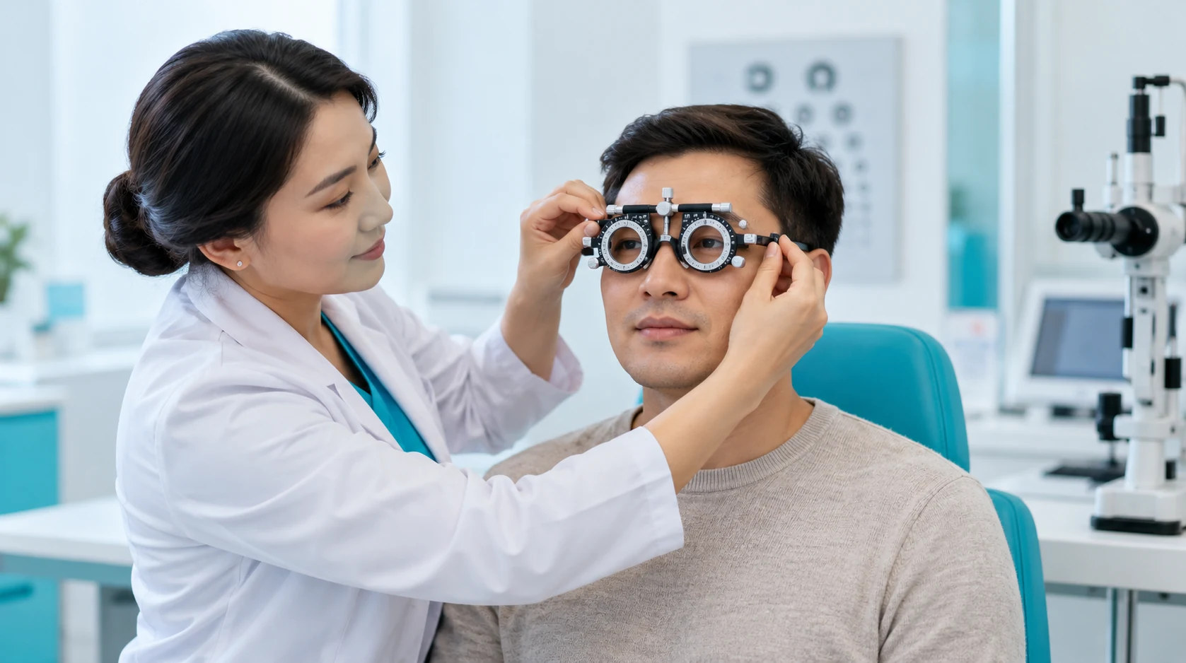
The doctor assesses the anterior segment of the eye and the measurement results, then determines whether a dilated fundus examination or specialized diagnostics is needed.
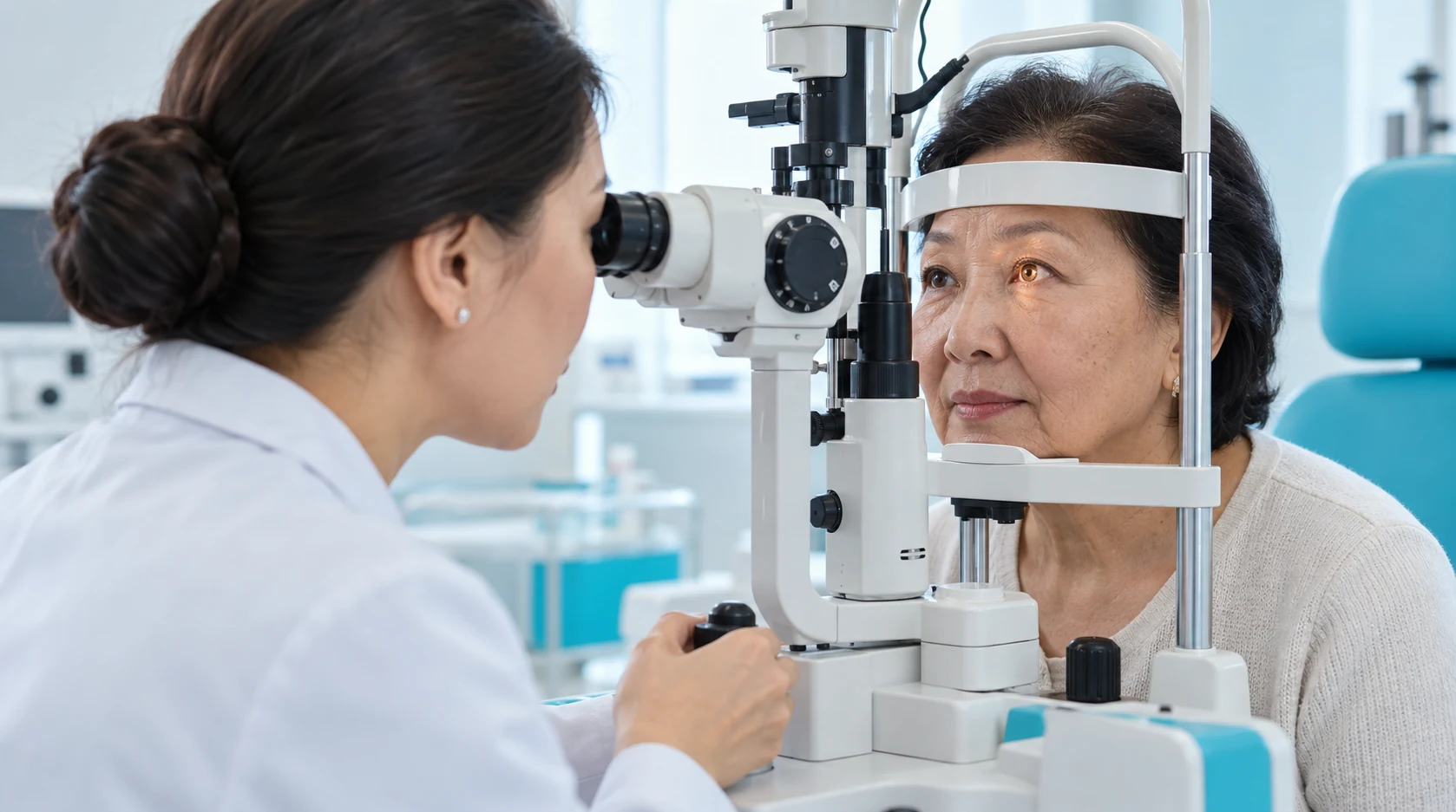
OCT, perimetry, topography, pachymetry, gonioscopy, ultrasound and other tests are prescribed to answer specific clinical questions, not for everyone.
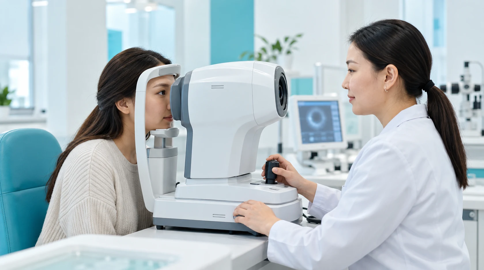
The ophthalmologist explains the findings and recommends monitoring, correction, treatment, further consultation or surgical preparation as needed.
Vision, eye health and symptoms are assessed together to understand what has changed.
The ophthalmologist explains the measurements and which findings need attention.
Additional tests follow symptoms and clinical indications rather than a standard package for everyone.
Results help the doctor decide on correction, treatment, monitoring or specialist referral.
To detect chronic intraocular pressure (high or low)
Learn morePrescribed when the ophthalmologist suspects structural changes in the deep layers of the eye.
Learn moreExamination of central and peripheral visual fields. This method is used to check for narrowing of the visible space.
Learn moreAnatomic Parameters and Internal Eye Structures
Learn moreIf your vision has changed, you have new symptoms, or you want to check your eye health, start with an ophthalmologist consultation.
The basic package includes: • Tonometry • Refractometry • Ocular ultrasound
Diagnostic Package for MSEK (Medical and Social Expert Commission) includes the following diagnostic examinations: • Ocular ultrasound (A- and B-scan) • Intraocular pressure (IOP) measurement • Refractometry • Pattern visual evoked potentials (VEP) using a checkerboard pattern • Flash visual evoked potentials (VEP) • Approximate visual acuity assessment using a checkerboard pattern • Static computerized perimetry
A team of experienced doctors across specialties.

Doctor of Medical Sciences, Associate Professor; Highest-category ophthalmologist

Candidate of Medical Sciences; Highest-category ophthalmologist


Highest-category ophthalmologist
A comprehensive eye evaluation starts with your symptoms and visual function, but it does not end with reading an eye chart. The doctor may assess refraction, intraocular pressure, the anterior segment and the fundus, and may order OCT, visual field testing, corneal topography, ultrasound or other tests when needed. The key value of the consultation is clinical interpretation.
The same complaint of “blurred vision” can have different causes involving refractive error, cataract, the cornea, retina, optic nerve or other structures. The ophthalmologist determines which tests are relevant and what the next step should be.
After basic measurements, the ophthalmologist analyses symptoms and findings, requests further tests if needed and provides a clear plan.
Testing depends on the visit's purpose: prevention, visual loss, correction fitting, surgical preparation and monitoring glaucoma, cataract or other eye conditions require different tests.
Tests are selected individually. Not all of the examinations listed are included in a single appointment.
Visual function and basic eye measurements are assessed. Confirm the exact examination contents with the clinic.
Used when needed to improve the view of internal eye structures. Temporary near blur and light sensitivity may follow.
Ordered when detailed cross-sectional imaging of the macula, retina or optic nerve is clinically useful.
Used in glaucoma assessment, selected neurologic situations and monitoring of specific eye diseases.
May be required for corneal disease assessment, refractive surgery planning and other indications.
Used when anatomical measurements or imaging of internal structures is required.
After diagnostics, patients may be referred to a glaucoma specialist, retina specialist, ophthalmic surgeon or another subspecialist.
The outcome of an examination is more than a printout of measurements. The patient should understand what was found, which changes need monitoring, whether additional tests or treatment are needed and when to return. The format for providing a written conclusion, test results and a next-step plan should be checked with the clinic.
Kazakhstan’s first specialized ophthalmology institute, operating since 1933.
Modern diagnostics and surgery using world-class equipment.
Leading ophthalmologists in Kazakhstan, scientists and experienced surgeons.
More than 30,000 operations and around 270,000 consultations annually.


Kazakhstan’s first specialized ophthalmology institute, operating since 1933.
Modern diagnostics and surgery using world-class equipment.
Leading ophthalmologists in Kazakhstan, scientists and experienced surgeons.
More than 30,000 operations and around 270,000 consultations annually.
Kazakhstan’s first specialized ophthalmology institute, operating since 1933.
Modern diagnostics and surgery using world-class equipment.
Leading ophthalmologists in Kazakhstan, scientists and experienced surgeons.
More than 30,000 operations and around 270,000 consultations annually.


