

Retinal laser photocoagulation
Laser photocoagulation creates controlled treatment spots in selected retinal areas.
Learn moreLaser photocoagulation creates controlled treatment spots in selected retinal areas.
Learn moreVitrectomy is microsurgery involving the vitreous and retina.
Learn moreRetinal treatment depends on the diagnosis and the condition of the macula and peripheral retina. Assessment determines whether monitoring, laser treatment, injections or surgery is appropriate.
Patients with retinal tears or symptomatic high-risk peripheral retinal lesions after dilated examination.
Patients with diabetic retinopathy or diabetic macular oedema.
Patients with wet AMD or another form of choroidal neovascularisation.
Patients with macular oedema due to retinal vein occlusion.
Patients with retinal detachment, vitreous haemorrhage or tractional retinal disease.
Patients with macular hole, epiretinal membrane or vitreomacular traction.
Patients after eye trauma or previous vitreoretinal surgery who need reassessment.
Not every peripheral retinal lesion needs prophylactic laser, and not every macular oedema is treated with the same injection.
With retinal detachment, timing of assessment can affect the functional prognosis.
Do not stop anticoagulants, antiplatelet medicines or systemic treatment on your own.
Urgent review is required for a sudden curtain, abrupt vision loss, flashes with many new floaters, eye trauma or severe pain.
Do not delay examination if you develop flashes, a sudden increase in floaters, a dark curtain, distorted straight lines, a central visual spot or sudden visual loss.
Bring OCT/OCT-A, ultrasound and angiography results, records of previous operations and a list of all medicines.
If you have diabetes, recent glucose and HbA1c results are useful.
Not every peripheral retinal lesion needs prophylactic laser, and not every macular oedema is treated with the same injection. With retinal detachment, timing of assessment can affect the functional prognosis. Urgent review is required for a sudden curtain, abrupt vision loss, flashes with many new floaters, eye trauma or severe pain. After an intravitreal injection or surgery, increasing pain, marked redness, sudden visual deterioration or purulent discharge requires urgent assessment.
Book a consultation
Consultation with a retina specialist/vitreoretinal surgeon, including symptom timing, diabetes, vascular risks and previous treatment.

Dilated retinal examination and core ophthalmic diagnostics.
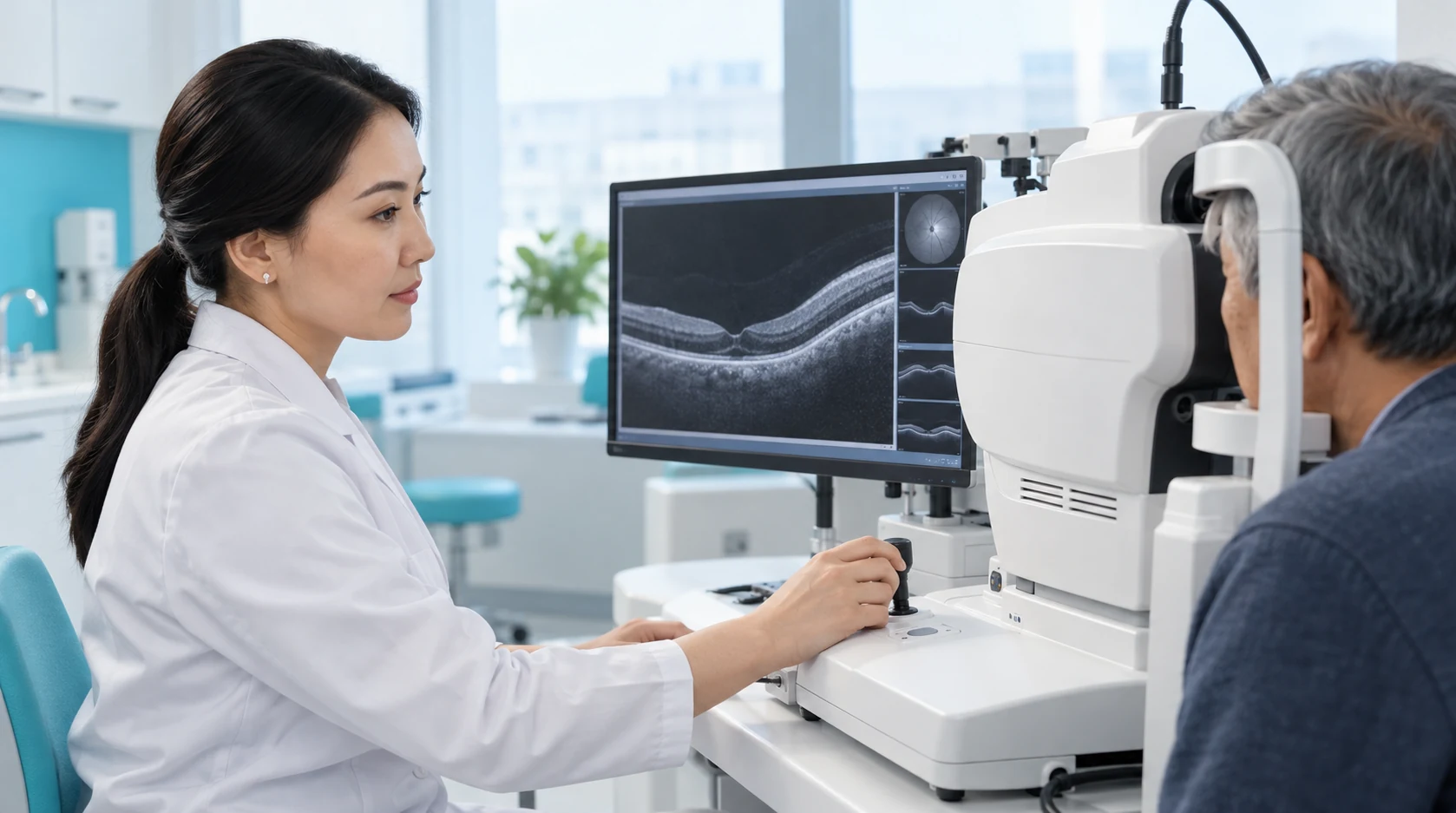
According to indications and availability.
Preventing detachment, reducing macular oedema, controlling neovascularisation, releasing traction or restoring retinal attachment.

Observation, laser, intravitreal therapy, vitrectomy, scleral intervention or a combination.
Follow-up on an individual schedule; chronic retinal disease may require a series of treatments and long-term monitoring.
The approach is selected according to the diagnosis, location of changes and condition of the central and peripheral retina.
Depending on the condition, laser, medication and surgical treatments may be used.
Timely treatment aims to preserve available visual function and reduce the risk of further deterioration.
Follow-up examinations help assess the retinal response to treatment and identify the need for further stages in time.
Colour applies only to the individual criterion: green = more favourable; yellow = situation dependent; red = limitation; grey = neutral/not directly comparable. It is not an overall ranking.
Colour applies only to the individual criterion: green = more favourable; yellow = situation dependent; red = limitation; grey = neutral/not directly comparable. It is not an overall ranking.
Book a consultation with a KazRIED retina specialist or vitreoretinal surgeon. After diagnostics, the doctor will determine whether observation, laser photocoagulation, intravitreal therapy, vitrectomy or another treatment route is appropriate.
A team of experienced doctors across specialties.

Doctor of Medical Sciences, Associate Professor; Highest-category ophthalmologist

Doctor of Medical Sciences; Highest-category ophthalmologist
Candidate of Medical Sciences, Associate Professor; Highest-category ophthalmologist

Candidate of Medical Sciences; Highest-category ophthalmologist

Candidate of Medical Sciences; Highest-category ophthalmologist

Candidate of Medical Sciences; Highest-category ophthalmologist

Doctor of Philosophy (PhD)
Master’s Student in Public Health

Highest-category ophthalmologist

Highest-category ophthalmologist
The retina responds differently to different diseases, so there is no single universal retinal treatment. Laser treatment, intravitreal therapy, and vitreoretinal surgery serve different purposes.
For a retinal tear without extensive detachment, the clinician may create a laser barrier around the area at risk. Laser photocoagulation may also be used for selected peripheral retinal degenerations when indicated.
Panretinal photocoagulation may be required for diabetic retinopathy or ischemic vascular disease.
Intravitreal medications are used when indicated for wet age-related macular degeneration (AMD), diabetic macular edema, and macular edema following retinal vein occlusion.
Vitrectomy may be required for retinal detachment, vitreous hemorrhage, an epiretinal membrane, vitreomacular traction, or a macular hole.
Depending on the diagnosis, surgery may include endolaser treatment, membrane peeling, and temporary tamponade. Gas, perfluorocarbon liquids, and silicone oil are used at different stages and for different durations according to the surgical plan.
The choice is based on a fundus examination, OCT, ultrasound, and other tests when indicated.
Treatment may aim to:
The visual prognosis also depends on macular condition, disease duration, ischemia, and coexisting conditions. Treatment goals are therefore established after a diagnostic assessment.
The follow-up plan depends on the diagnosis and treatment performed:
In diabetic and vascular disease, managing the underlying systemic condition remains part of preserving vision.
Kazakhstan’s first specialized ophthalmology institute, operating since 1933.
Modern diagnostics and surgery using world-class equipment.
Leading ophthalmologists in Kazakhstan, scientists and experienced surgeons.
More than 30,000 operations and around 270,000 consultations annually.


Kazakhstan’s first specialized ophthalmology institute, operating since 1933.
Modern diagnostics and surgery using world-class equipment.
Leading ophthalmologists in Kazakhstan, scientists and experienced surgeons.
More than 30,000 operations and around 270,000 consultations annually.
Kazakhstan’s first specialized ophthalmology institute, operating since 1933.
Modern diagnostics and surgery using world-class equipment.
Leading ophthalmologists in Kazakhstan, scientists and experienced surgeons.
More than 30,000 operations and around 270,000 consultations annually.


