

Posterior capsule opacification
Slit-lamp examination shows posterior capsule opacification involving the central visual axis.
Posterior capsule opacification is clouding behind an artificial lens after cataract surgery. When indicated, an Nd:YAG laser creates an opening in the capsule to clear the visual axis.
Slit-lamp examination shows posterior capsule opacification involving the central visual axis.
Glare, halos or reduced contrast developed after IOL implantation and interfere with daily activities.
Visual acuity has declined and the ophthalmologist considers the degree of PCO consistent with the symptoms.
The anterior and posterior capsules, IOL position and retinal condition allow YAG capsulotomy to be considered safely.
The patient needs differential diagnosis between PCO, retinal disease, glaucoma and IOL-related problems after cataract surgery.
Vision initially improved after cataract surgery but later became progressively cloudy again.
Nd:YAG capsulotomy is a minimally invasive laser procedure but it is not risk-free. Possible events include a temporary intraocular-pressure rise, inflammation, new floaters, laser pitting of the IOL, macular oedema and rarer complications such as retinal tear or detachment.
Additional caution is needed in glaucoma or ocular hypertension, high myopia, previous retinal tears or detachment, uveitis, macular disease, unstable IOL position and when future IOL rotation or exchange may be required.
If vision became cloudy again after successful cataract surgery, or glare, halos or reduced contrast returned, arrange an eye examination. Bring the previous operative report and, if available, information about the implanted IOL, retinal disease, glaucoma , high myopia , uveitis and prior laser procedures.
Do not use eye drops marketed to “dissolve” secondary cataract without medical advice because drops do not create an optical opening in an opaque posterior capsule.
Seek urgent eye care for sudden visual loss, severe pain, marked redness, flashes, a curtain-like shadow or a sudden increase in floaters. Sudden visual loss, flashes, a curtain-like shadow, severe pain or many new floaters require urgent assessment rather than routine YAG treatment.
Nd:YAG capsulotomy is a minimally invasive laser procedure but it is not risk-free.
Book a consultation
The ophthalmologist reviews the date and course of the previous cataract operation and asks about glaucoma, high myopia, uveitis and retinal disease.

Visual acuity and intraocular pressure are checked, and IOL position and the posterior capsule are assessed by slit-lamp biomicroscopy; the fundus and OCT are assessed when indicated.
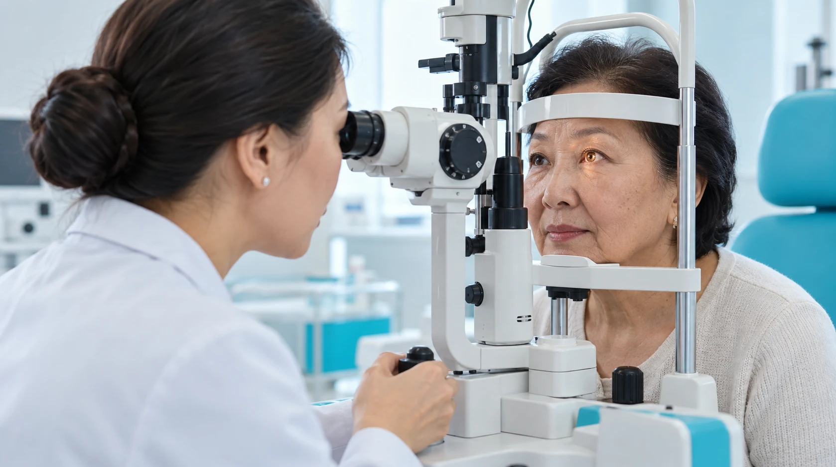
If PCO is clinically significant and there are no contraindications, the doctor plans Nd:YAG laser capsulotomy and explains the expected effect and risks.

Dilating drops and other topical medication may be used according to local protocol; the exact KazRIED regimen requires confirmation.

Laser pulses create a central opening in the opaque posterior capsule using sufficient but not excessive energy.
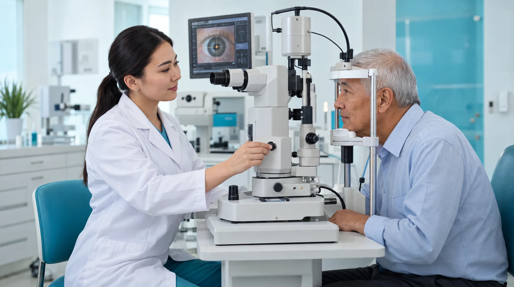
After treatment the physician individualises IOP monitoring, anti-inflammatory medication and follow-up according to risk factors.

The laser creates an opening in the cloudy posterior capsule when it obstructs the passage of light.
Treatment of posterior capsule opacification usually clears the capsule without replacing the artificial lens.
Standard laser posterior capsulotomy does not require a corneal incision or sutures.
When capsule clouding causes reduced vision, restoring the passage of light can improve image clarity.
If vision has declined again after cataract surgery, book an examination at KazRIED. The doctor will assess the posterior capsule, IOL, intraocular pressure and fundus and determine whether YAG laser capsulotomy is appropriate or whether another cause needs treatment.
A team of experienced doctors across specialties.

Candidate of Medical Sciences; Highest-category ophthalmologist
Highest-category ophthalmologist
Highest-category ophthalmologist

PhD Student

After phacoemulsification, the cloudy natural lens has been removed, and the artificial lens usually remains inside the preserved capsular bag.
Residual lens epithelial cells may migrate and proliferate on the posterior capsule, causing fibrosis, folds, or formations known as Elschnig pearls.
If these changes affect the central optical zone, the patient may again notice hazy vision, reduced visual acuity, glare, halos, and reduced contrast.
This condition is called posterior capsule opacification (PCO), commonly referred to as a secondary cataract. It does not mean that the original cataract has “returned,” nor does it automatically indicate a problem with the IOL.
Before laser treatment, the clinician must confirm that the symptoms are caused by capsule opacification rather than a condition affecting the macula, retina, or optic nerve, elevated intraocular pressure, clouding of the IOL itself, or another cause.
For clinically significant PCO in adults, the standard treatment is Nd:YAG laser posterior capsulotomy. Laser pulses create a central opening in the cloudy capsule.
IOL replacement is usually unnecessary if the artificial lens is clear, correctly positioned, and there are no other indications for intraocular surgery.
Typical PCO: gradual visual decline after initially successful surgery, central posterior-capsule opacity and stable IOL – consider YAG when indicated.
PCO + glaucoma: assess baseline IOP and optic nerve; pressure prophylaxis and monitoring are individualised.
PCO + high myopia / retinal history: evaluate peripheral retina and symptoms of flashes/floaters more carefully before YAG.
PCO + macular disease: OCT helps estimate how much visual loss is capsular and what laser treatment can reasonably improve.
Suspected IOL opacity or dislocation: do not perform YAG automatically; first determine whether IOL exchange or repositioning may be needed.
Dense fibrotic membrane or unusual anatomy: the laser surgeon decides whether YAG is sufficient or a surgical approach is preferable.
Consultation with Candidate of Medical Sciences: When booked with a physician of this category
Highest-category / >10 years consultation: Depends on the selected specialist
Biomicroscopy: Examination of posterior capsule and anterior segment
Ophthalmoscopy: When indicated to assess the fundus
Cirrus-HD macular OCT: When indicated, if the reduced vision is not fully explained by the posterior capsule findings.
Spectralis posterior-segment OCT: Alternative OCT when prescribed
Kazakhstan’s first specialized ophthalmology institute, operating since 1933.
Modern diagnostics and surgery using world-class equipment.
Leading ophthalmologists in Kazakhstan, scientists and experienced surgeons.
More than 30,000 operations and around 270,000 consultations annually.


Kazakhstan’s first specialized ophthalmology institute, operating since 1933.
Modern diagnostics and surgery using world-class equipment.
Leading ophthalmologists in Kazakhstan, scientists and experienced surgeons.
More than 30,000 operations and around 270,000 consultations annually.
Kazakhstan’s first specialized ophthalmology institute, operating since 1933.
Modern diagnostics and surgery using world-class equipment.
Leading ophthalmologists in Kazakhstan, scientists and experienced surgeons.
More than 30,000 operations and around 270,000 consultations annually.


