
Tonometry
Tonometry measures intraocular pressure using a contact or non-contact method.
Learn moreTonometry measures intraocular pressure using a contact or non-contact method.
Learn morePachymetry measures corneal thickness.
Learn moreOCT produces cross-sectional images of eye structures without surgery.
Learn morePerimetry tests the visual field and helps identify areas of reduced sensitivity.
Learn moreGlaucoma assessment evaluates eye pressure, the optic nerve and visual fields. The combined findings help confirm the diagnosis, establish the stage and plan follow-up.
IOP has been elevated at least once or there is a meaningful pressure asymmetry between the eyes.
The optic disc appears suspicious, with increased cupping, rim thinning or other concerning changes.
There is a family history of glaucoma, especially together with other risk factors.
High myopia, marked hyperopia, pseudoexfoliation, diabetes, previous eye trauma or surgery increases the need for targeted assessment.
Steroid medicines have been used for a prolonged period in drops, inhalers, tablets or injections.
Glaucoma is already diagnosed and stability needs to be assessed with visual fields, OCT and IOP measurements.
There are episodes of pain, halos or blurred vision, particularly when a narrow anterior chamber angle is suspected.
Bring previous IOP readings, OCT and visual-field reports, surgical records and a list of all medicines, including eye drops and steroids. Do not stop glaucoma drops before testing unless instructed by your doctor. If you are already under glaucoma follow-up, older tests are valuable for progression analysis.
Tell the physician about family history, previous eye trauma or surgery, high myopia or hyperopia , diabetes and long-term corticosteroid use.
Severe eye pain, marked redness, sudden blurred vision, coloured halos, headache, nausea or vomiting can be signs of acute angle closure and require urgent ophthalmic assessment rather than a routine appointment. A normal single IOP reading does not exclude glaucoma, and elevated IOP alone is not the same as a glaucoma diagnosis.
Severe eye pain, marked redness, sudden blurred vision, coloured halos, headache, nausea or vomiting can be signs of acute angle closure and require urgent ophthalmic assessment rather than a routine appointment. A normal single IOP reading does not exclude glaucoma, and elevated IOP alone is not the same as a glaucoma diagnosis.
Book an eye examination
Family history, medicines, surgery, injuries, refraction and associated conditions.

Basic vision and anterior-segment assessment.

Pressure is measured by the selected method; repeat or diurnal measurements are arranged when needed.
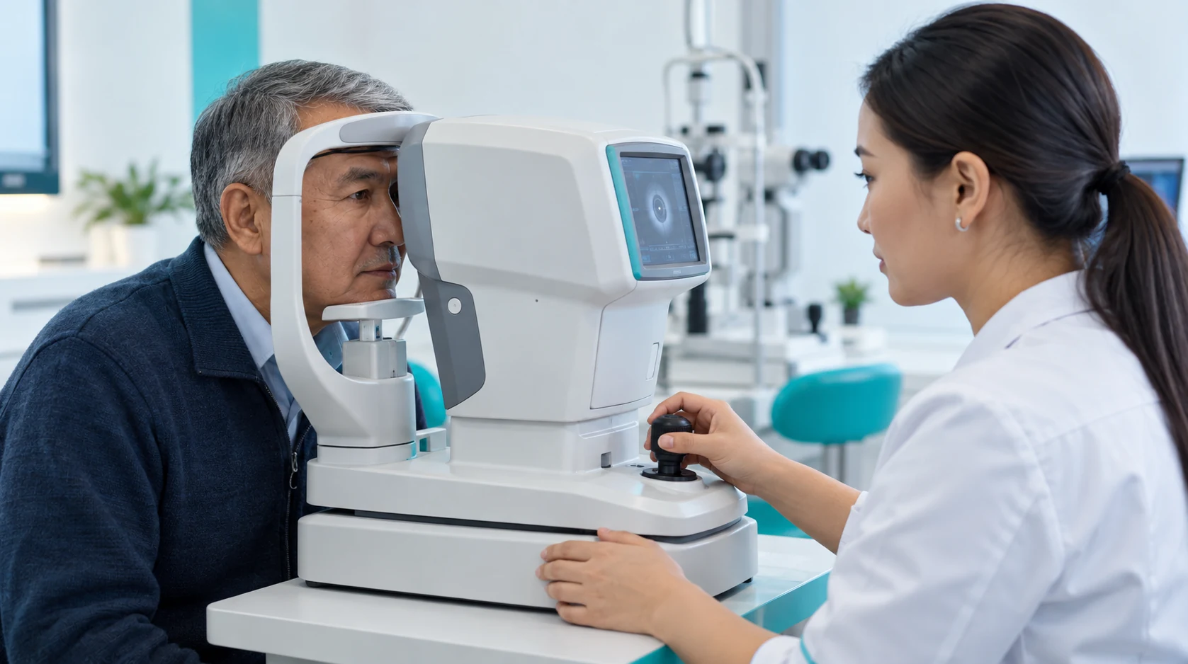
Corneal thickness is considered when interpreting pressure and risk.
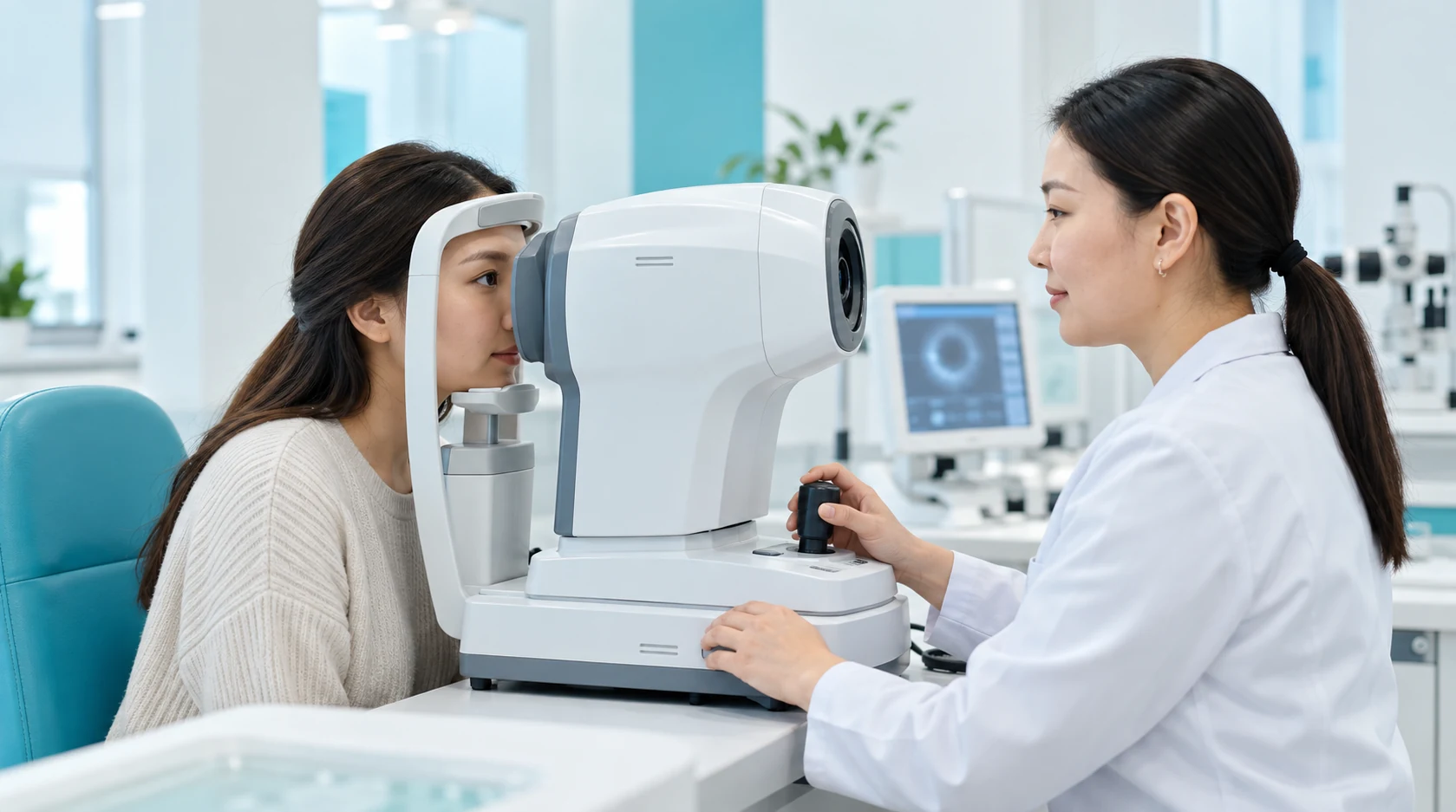
The doctor assesses the anterior-chamber angle before establishing the glaucoma mechanism.
Lens-assisted biomicroscopy, photography or HRT when indicated.
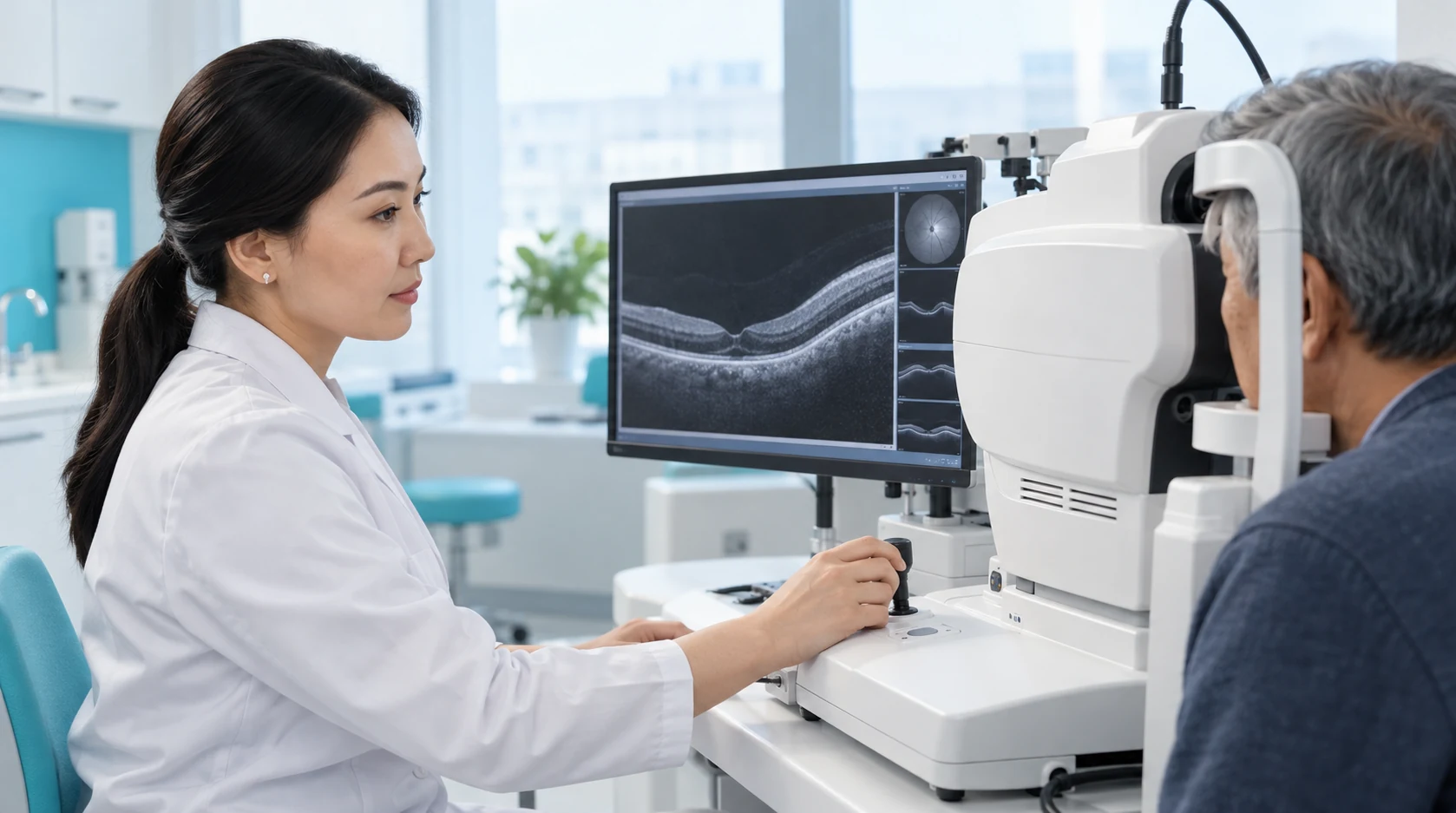
Nerve fibre structure and comparison with the normative database are assessed, considering artefacts.
Visual-field function and defects are assessed; doubtful results require repeat testing.
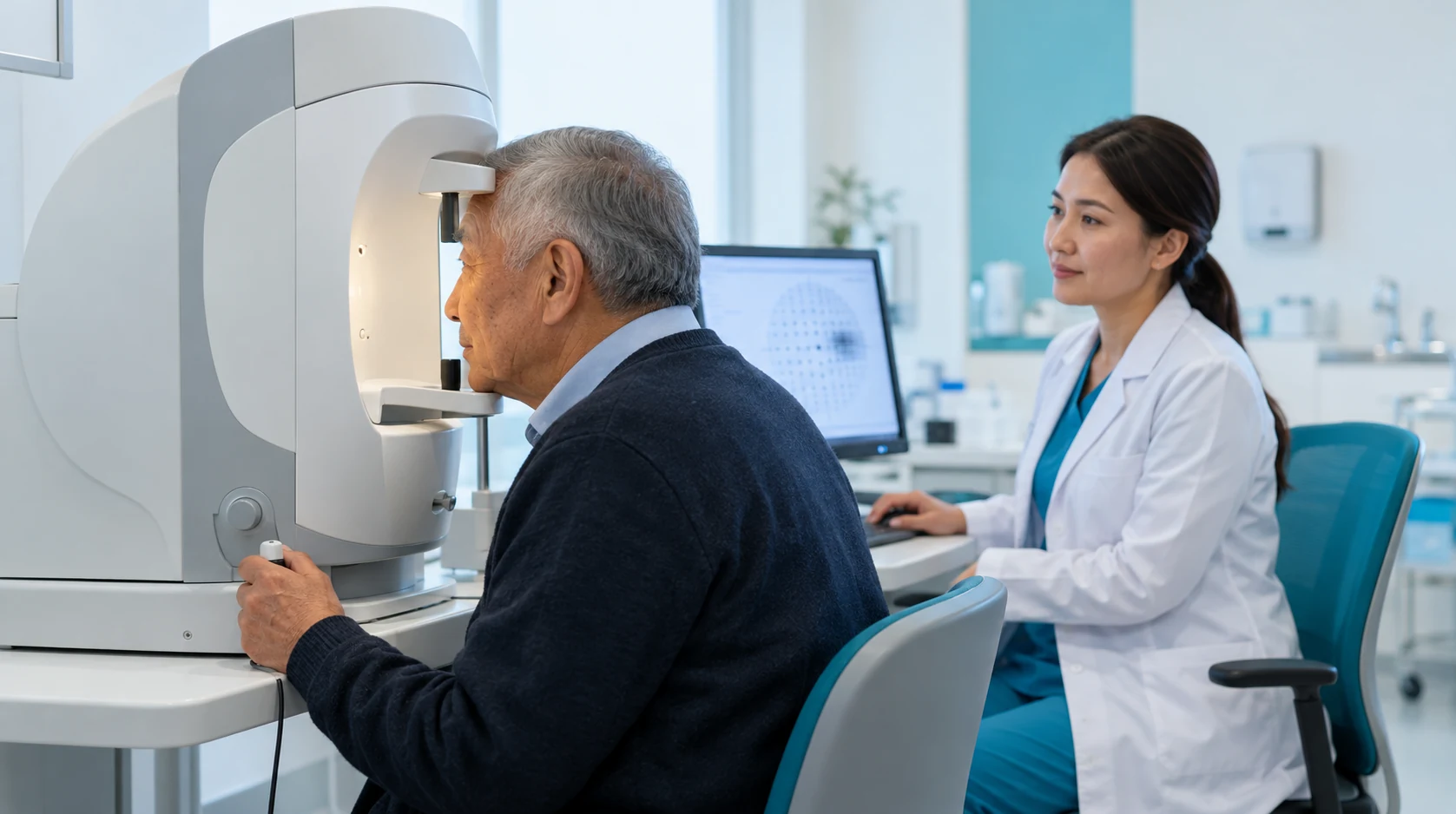
Glaucoma, suspected glaucoma, ocular hypertension or another diagnosis; treatment needs and follow-up are determined.
Comprehensive assessment can detect suspicious changes before a person notices deterioration.
Tonometry provides an intraocular-pressure reading for diagnosis and follow-up.
Imaging helps identify structural changes relevant to glaucoma diagnosis.
Perimetry shows how a person detects objects in central and peripheral vision.
If IOP has been elevated, the optic nerve looks suspicious, there is a family history of glaucoma or you already require glaucoma follow-up. The physician will select the appropriate tests and interpret pressure, optic nerve structure and visual-field function together.
A team of experienced doctors across specialties.

Candidate of Medical Sciences; Highest-category ophthalmologist

Candidate of Medical Sciences; Highest-category ophthalmologist

Candidate of Medical Sciences; Highest-category ophthalmologist

Highest-category ophthalmologist

Highest-category ophthalmologist

Highest-category ophthalmologist
Highest-category ophthalmologist

Highest-category ophthalmologist
Highest-category ophthalmologist

Highest-category ophthalmologist
Glaucoma is diagnosed using a combination of structural and functional findings. Elevated intraocular pressure (IOP) is an important risk factor, but it is not a requirement for the disease.
In normal-tension glaucoma, optic nerve damage may progress at IOP levels that do not appear elevated during a single measurement.
Elevated pressure without characteristic optic nerve damage or visual field loss may indicate ocular hypertension.
When necessary, additional testing may include repeated pressure measurements, tonography, Heidelberg retinal tomography (HRT), optic disc photography, ultrasound biomicroscopy (UBM), or anterior segment OCT.
Comparing findings over time is particularly important for establishing the diagnosis and assessing progression. A single test should not be interpreted in isolation from the other examination findings.
Repeat assessment is needed to confirm the diagnosis and evaluate disease progression.
The frequency of monitoring depends on the glaucoma stage, risk level, stability of intraocular pressure (IOP), and the rate of change in visual field and OCT findings.
When angle-closure glaucoma is suspected, gonioscopy and anterior segment imaging are particularly important. In high myopia, OCT findings require especially cautious interpretation.
The “Glaucoma Diagnostics” package: individual tests do not automatically constitute a complete diagnostic package.
Глаукома ұзақ уақыт бойы айқын шағымдар туғызбауы мүмкін. Сондықтан диагностиканың мақсаты — көру өрісі едәуір тарылғанға дейін зақымдануды анықтау және кейінгі салыстыру үшін бастапқы көрсеткіштерді тіркеу.
Пациентке әр кезеңдегі ОКТ және периметрия нәтижелерін сақтаған пайдалы. Зерттеулердің салыстыруға келуі дәрігерге аурудың шынайы үдеуін тест нәтижелерінің құбылмалылығынан ажыратуға көмектеседі.
Жекелеген зерттеулердің нәтижелері сәйкес келмесе, бұл тестілердің бірі «дұрыс емес» дегенді білдірмейді.
Ерте кезеңдерде ОКТ-дағы құрылымдық өзгерістер көру өрісінің ақауларынан бұрын пайда болуы мүмкін. Ал басқа жағдайларда функционалдық өзгерістер айқынырақ байқалады.
Шешімді офтальмолог кешенді бағалаудан кейін қабылдайды.
Kazakhstan’s first specialized ophthalmology institute, operating since 1933.
Modern diagnostics and surgery using world-class equipment.
Leading ophthalmologists in Kazakhstan, scientists and experienced surgeons.
More than 30,000 operations and around 270,000 consultations annually.


Kazakhstan’s first specialized ophthalmology institute, operating since 1933.
Modern diagnostics and surgery using world-class equipment.
Leading ophthalmologists in Kazakhstan, scientists and experienced surgeons.
More than 30,000 operations and around 270,000 consultations annually.
Kazakhstan’s first specialized ophthalmology institute, operating since 1933.
Modern diagnostics and surgery using world-class equipment.
Leading ophthalmologists in Kazakhstan, scientists and experienced surgeons.
More than 30,000 operations and around 270,000 consultations annually.


